

Chest wall malformations (CWM) can be exhibited in several ways and can cause both functional respiratory impairment and psychosocial concerns including aversion to sports and public exposure. CWMs occur quite frequently, however they are often misdiagnosed or not diagnosed at all, leading to a continued progression of the malformation.
There are five different classifications of CWM types that are dependant on origin site of the malformation. These include: cartilaginous, costal, chondro-costal, sternal, and clavicle-scapular. It is important to determine the correct classification of CWM to ensure the proper treatment is provided to the patient.
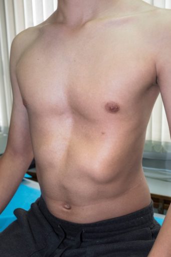
The first classification, cartilaginous, contains two common types of malformations: pectus excavatum (PE) and pectus carinatum (PC). PE is the most commonly-occuring thoracic malformation and is characterized by a deep sternal depression. Patients with PE are considered for surgery if they have two or more symptoms, including: progression of the malformation, cardiac compression, cava vein or pulmonary compression, and significant body image disturbance. If approved for surgery, Nuss procedure has the highest satisfaction rates. The Nuss procedure utilizes a rotated metal bar to correct the deformity.
PC occurs at a rate of five times less than PE and is predominantly observed in male patients. It is characterized by asymmetry along different structures surrounding the chest. Possible treatment for PC patients include surgeries such as: costal excision surgery, sternal osteotomy, intrathoracic compression procedure, thoracoscopic cartilage resection, and mini-invasive submuscular dissection.
The second classification, costal, pertains to malformed cartilage that causes a unilateral or bilateral depression in the thoracic wall. The most common treatment for this type of CWM is cartilage excision.
The third classification, chondrocostal, is associated with Poland Syndrome which is the absence or hypoplasia of the pectoralis major muscle. The absence of the pectoralis major muscle can also cause deformities of the chest wall, breast, and upper limb. Thoracoplasty is a common surgical procedure used to correct this issue and is sometimes used with other surgical procedures such as: latissimus dorsi or rectal abdominal muscle transposition, lipofilling, or omental flap technique.
The fourth classification, sternal, is defined by a sternal cleft, Currarino syndrome, or Pouter Pigeon Breast. Sternal clefts can be partial or complete and often times they are associated with other defects such as maxillofacial mehmangiomas, cleft lip or cleft palate, pectus excavatum, connectival nevi, supraumbilical raphe, gastroschisis, cardiac defects, aortic coarctation, eye abnormalities, posterior fossa anomalies, and hidden haemangiomas. Surgical intervention is recommended from the neonatal period through the first months of life. Otherwise, more invasive surgery may be needed including: sternoclavicular disarticulation, sternal isolation, inferior sternal osteotomy and medialization of the neck muscles after separation of their sternoclavicular attachments laterally.
The fifth and final classification is clavicle-scapular results from deformities in the clavicle region or scapular region that subsequently cause the chest wall to not sit or function correctly. This type of CWM is not as commonly seen and, therefore, surgical intervention is usually decided on a case by case basis.