The Borstein Family Foundation Melanoma Program: A Center of Excellence

The Borstein Family Foundation Melanoma Program at Saint John's Cancer Institute brings together specialists in melanoma, skin cancer, and soft tissue tumors to provide individualized, multidisciplinary care. Our approach combines clinical evaluation, advanced treatment options, and a longstanding commitment to cancer research. Working closely with Providence Saint John's Health Center, our physicians and researchers help patients understand their diagnosis, evaluate appropriate treatments, and access coordinated care supported by scientific discovery.

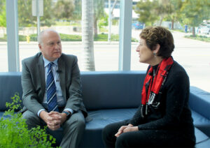
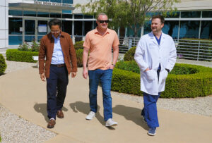
Saint John's Melanoma Team

The Borstein Family Foundation Melanoma Program

At Saint John’s Cancer Institute, melanoma care brings together surgical oncology, medical oncology, dermatology, pathology, radiation oncology, and clinical research to address the needs of patients with melanoma, other skin cancers, and soft tissue tumors. The Borstein Family Foundation Melanoma Program supports an approach centered on timely evaluation, accurate diagnosis, individualized treatment planning, and coordinated care.

A History of Innovation and Collaboration

Dr. Donald L. Morton
Dr. Donald Morton, Saint John’s Cancer Institute Co-founder and pioneer of immunotherapy and the sentinel lymph node discention procedure.

The Institute’s melanoma research history is closely associated with Donald L. Morton, MD, whose investigations into cancer immunotherapy and sentinel lymph node mapping helped shape modern melanoma care. His work and subsequent collaborative research contributed to advances in understanding how melanoma spreads, how regional lymph nodes should be evaluated, and when extensive lymph node surgery may be avoided.

Research at the former John Wayne Cancer Institute, now Saint John’s Cancer Institute, helped establish sentinel lymph node biopsy as an important staging procedure. Later clinical investigations, including the international Multicenter Selective Lymphadenectomy Trials, provided evidence that has continued to refine melanoma surgical decisions.

Support from the Borstein Family Foundation helps sustain a program that connects this research history with clinical care and investigation into evolving treatments for melanoma and other complex cancers.

Connecting Clinical Research With Treatment

The Institute maintains melanoma research resources, including an established collection of tumor specimens and associated clinical information used to investigate cancer biology, recurrence, prognosis, and treatment response. These resources support studies intended to improve how melanoma is understood and treated.

Collaborative research also examines how melanoma interacts with the immune system, how molecular changes influence cancer behavior, and which treatment approaches may benefit particular groups of patients. Findings from clinical and laboratory research can inform the development and evaluation of future therapies.

For patients, these efforts complement the clinical work of specialists who evaluate skin lesions, establish diagnoses, determine disease stage, and recommend appropriate treatment options. Research participation and available clinical trials are considered according to individual eligibility and clinical circumstances.

Explore the Institute’s Melanoma Research Program and Database or visit Clinical Trials to learn more about current research.

Melanoma, Skin Cancer and Soft Tissue Tumor Conditions

Melanoma, other skin cancers, and soft tissue sarcomas differ considerably in how they develop, appear, and behave. Some are commonly identified through visible skin changes, while others develop beneath the skin and may initially be noticed as a lump or swelling. Accurate diagnosis helps determine whether treatment is needed and which specialists should be involved.

The Borstein Family Foundation Melanoma Program provides evaluation and coordinated care for patients with the following conditions. Explore each page for more information about symptoms, risk factors, diagnosis, staging, and treatment.

Basal Cell Carcinoma

Clinical image illustrating basal cell carcinoma of the skin
Basal cell carcinoma can develop as a persistent or changing skin lesion.

Basal cell carcinoma is the most common type of skin cancer. It develops from cells in the skin’s basal layer and often occurs on areas exposed to ultraviolet radiation, including the face, neck, and arms. Most cases grow slowly, but untreated tumors can damage surrounding tissues.

Common features include:

  • A shiny or pearly bump, persistent pink growth, or scaly patch.
  • A sore that does not heal or repeatedly bleeds and crusts.
  • A scar-like area or changing lesion, sometimes with little discomfort.

Learn more about Basal Cell Carcinoma.

Melanoma

Clinical image illustrating melanoma skin cancer
Melanoma may develop within an existing mole or appear as a new or changing skin lesion.

Melanoma develops from melanocytes, the cells that produce skin pigment. It can arise on sun-exposed or less-exposed areas of the body and may spread to lymph nodes or distant organs if not identified and treated appropriately. Early diagnosis is important because many localized melanomas can be treated successfully with surgery.

Possible warning signs include:

  • A new mole or an existing spot that changes in size, shape, or color.
  • An irregular border, uneven pigmentation, or a lesion unlike other moles.
  • Bleeding, itching, or another persistent change in a pigmented or nonpigmented lesion.

Learn more about Melanoma, including risk factors, the ABCDE warning signs, staging, and treatment.

Merkel Cell Carcinoma

Clinical image illustrating Merkel cell carcinoma
Merkel cell carcinoma may appear as a rapidly growing, often painless skin nodule.

Merkel cell carcinoma is an uncommon but potentially aggressive skin cancer. It frequently appears as a firm, painless, rapidly enlarging bump that may be pink, red, purple, or skin-colored. It occurs more often in older adults and people with weakened immune systems.

Important characteristics include:

  • A new or rapidly growing skin nodule, sometimes without other symptoms.
  • An association with ultraviolet exposure and, in many cases, Merkel cell polyomavirus.
  • A potential to spread to nearby lymph nodes or distant sites, making timely evaluation important.

Learn more about Merkel Cell Carcinoma.

Soft Tissue Sarcoma

Medical image representing soft tissue sarcoma
Soft tissue sarcomas can develop in the body’s connective and supporting tissues.

Soft tissue sarcomas are a diverse group of cancers that develop in tissues such as muscle, fat, blood vessels, nerves, and fibrous structures. Unlike most skin cancers, they may arise deep beneath the skin and can occur in the arms, legs, trunk, or other parts of the body.

Features requiring evaluation may include:

  • A new or enlarging lump, particularly one that is deep or difficult to move.
  • A swelling or mass that may initially be painless.
  • Pain, pressure, or changes in function when a tumor affects nearby structures.

Learn more about Soft Tissue Sarcoma, including diagnosis, staging, and treatment considerations.

Squamous Cell Carcinoma of the Skin

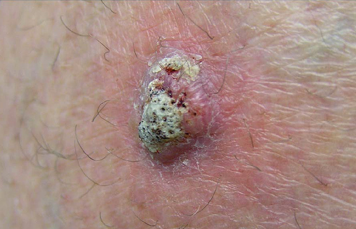
Clinical image illustrating cutaneous squamous cell carcinoma
Squamous cell carcinoma often appears as a persistent scaly, crusted, or growing skin lesion.

Cutaneous squamous cell carcinoma develops from squamous cells in the outer layers of the skin. It often occurs in areas with substantial ultraviolet exposure, such as the face, scalp, ears, and hands. Many cases are successfully treated when identified early, but some tumors may grow aggressively or spread.

Common features include:

  • A rough or scaly patch, firm bump, or thickened skin lesion.
  • A sore that bleeds, crusts, or fails to heal.
  • Greater concern for lesions that grow rapidly, recur, or develop in higher-risk locations.

Learn more about Squamous Cell Carcinoma of the Skin.

For a consolidated introduction to these diseases, visit our Conditions Overview.

Early Detection and Skin Cancer Screening

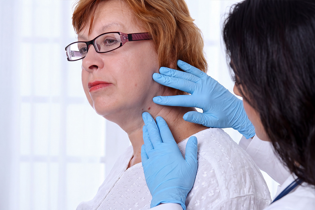
Skin lesion evaluation at the Pigmented Skin Lesion Clinic at Saint John's Health Center
The Pigmented Skin Lesion Clinic provides evaluation of suspicious moles and skin lesions in Santa Monica.

Melanoma and other skin cancers may begin with subtle changes that are difficult to recognize without clinical evaluation. A new or changing mole, persistent sore, unusual pigmentation, or other concerning skin lesion may warrant examination by a qualified healthcare professional.

Through the Pigmented Skin Lesion Clinic, Saint John’s Health Center provides specialized evaluation of suspicious pigmented lesions, with access to additional diagnostic or treatment resources when appropriate.

Early identification may allow skin cancer to be treated before it becomes more extensive. People with a personal or family history of melanoma, substantial ultraviolet exposure, numerous or atypical moles, or a changing lesion should discuss their individual evaluation and screening needs with a healthcare professional.

Learn more about Skin Cancer Screening and the Pigmented Skin Lesion Clinic.

Melanoma Treatment Options

Melanoma treatment depends on the cancer’s stage, pathology findings, molecular characteristics, location, and whether it has spread beyond the original site. Surgery is the primary treatment for most localized melanomas, while immunotherapy, targeted medication, radiation, and other approaches may be appropriate for selected patients.

Some procedures, including sentinel lymph node biopsy, are performed primarily to establish the stage of disease rather than directly treat the original tumor. Understanding these distinctions can help patients and families evaluate their options and participate in treatment decisions.

Chemotherapy

Chemotherapy uses medications that interfere with cancer cell growth and division. Although immunotherapy and targeted therapy have largely replaced conventional chemotherapy as preferred systemic options for many patients with advanced melanoma, chemotherapy may still be considered in selected situations when other treatments are unsuitable or have stopped working.

Learn more about Chemotherapy for Melanoma.

Completion Node Dissection

Completion lymph node dissection removes additional lymph nodes from a regional lymph node basin after melanoma has been identified in a sentinel node. Research has changed the procedure’s role: many patients with microscopic sentinel node involvement can undergo careful ultrasound surveillance instead of immediate additional surgery. More extensive lymph node removal remains appropriate in selected circumstances, including certain patients with clinically apparent regional disease.

Learn more about Completion Lymph Node Dissection.

Immunotherapy

Immunotherapy helps the immune system recognize and attack melanoma cells. Established treatments include immune checkpoint inhibitors, which may be used for advanced melanoma and in selected patients before or after surgery. Cellular immunotherapy and treatments delivered directly into tumors offer additional options for eligible patients.

Learn more about Immunotherapy for Melanoma, including the Institute’s history of immunotherapy research.

Radiation Therapy

Radiation therapy equipment used for targeted cancer treatment
Radiation therapy may help control selected melanoma tumors or relieve symptoms at specific sites.

Radiation therapy uses directed high-energy radiation to damage cancer cells within a defined treatment area. It is not routinely necessary for most localized melanomas, but may be considered for selected patients following surgery, when surgery is not feasible, or when melanoma has recurred or spread to particular locations.

At Saint John’s Health Center, Vasek Polak Radiation Oncology provides radiation treatment planning and specialized techniques in coordination with the patient’s cancer care team.

Learn more about Radiation Therapy for Melanoma.

Sentinel Lymph Node Biopsy

Illustration representing sentinel lymph node evaluation for melanoma
Research at Saint John’s Cancer Institute helped establish sentinel lymph node biopsy as an important melanoma staging procedure.

Sentinel lymph node biopsy identifies and removes selected lymph nodes that first receive drainage from the original melanoma site. The nodes are examined for microscopic cancer cells, providing information that helps establish the disease stage, estimate recurrence risk, and guide recommendations for additional treatment or surveillance.

Research by Donald L. Morton, MD, and colleagues helped develop this approach, allowing more selective evaluation of regional lymph nodes.

Learn more about Sentinel Lymph Node Biopsy.

Surgery (Wide Local Excision)

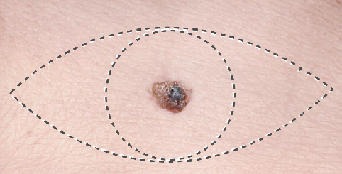
Medical illustration of wide local excision of a melanoma and surrounding tissue
Wide local excision removes the original melanoma site and an appropriate margin of surrounding skin.

Wide local excision is the primary treatment for most localized cutaneous melanomas. The surgeon removes the melanoma or original biopsy site along with an appropriate margin of normal-appearing tissue. The recommended margin depends largely on Breslow thickness and may be modified for particular anatomical or clinical circumstances.

Many patients with early-stage melanoma require no additional cancer-directed treatment after surgery, although lymph node evaluation or other therapies may be recommended according to the pathology findings.

Learn more about Surgery (Wide Local Excision).

Targeted Therapy

Targeted therapy uses medications designed to interfere with specific molecular changes that help melanoma cells grow. Certain melanomas contain BRAF mutations that may respond to combinations of BRAF and MEK inhibitors. Molecular testing helps determine whether these medications are appropriate for patients with advanced disease or selected higher-risk melanomas following surgery.

Research is also advancing personalized cancer treatments, including investigational mRNA vaccines designed around mutations found within an individual patient’s melanoma. These vaccines are a form of emerging immunotherapy, distinct from established molecularly targeted drugs.

Learn more about Targeted Therapy for Melanoma.

Research, Clinical Trials and Treatment Planning

Clinical trials play an important role in evaluating emerging melanoma treatments and improving how existing therapies are used. Research into immunotherapy, molecularly targeted medications, cellular therapies, and personalized cancer vaccines continues to shape the future of melanoma care.

At Saint John’s Cancer Institute, multidisciplinary evaluation can help patients understand established treatment options and whether a clinical trial may be appropriate. Study availability and eligibility depend on the specific research protocol and the patient’s clinical circumstances.

For a more complete explanation of these options, visit our Melanoma Treatment Overview. You can also explore the Institute’s Clinical Trials Program.

If you have questions regarding melanoma or other skin conditions, treatments, or clinical trials, please call today or click here to schedule an appointment or for an expert second opinion.

Safety and Efficacy of the Combination of Nivolumab Plus Ipilimumab in Patients with Melanoma and Asymptomatic or Symptomatic Brain Metastases