
Understanding Squamous Cell Carcinoma of the Skin
Squamous cell carcinoma (SCC) is the second most common form of skin cancer, developing from squamous cells in the outer layer of the skin. Often associated with years of ultraviolet (UV) exposure, SCC can appear as a rough patch, persistent sore, or raised growth. Most cases are highly treatable when identified early, but some tumors can invade deeper tissues or spread to nearby lymph nodes and other parts of the body.
Cutaneous squamous cell carcinoma can occur on any part of the skin, although it is frequently found on the face, ears, scalp, neck, arms, and hands. Risk increases with age and accumulated sun exposure, but SCC also develops in people with darker skin tones and in areas that receive little sunlight. Understanding new or changing lesions can help patients recognize when a professional skin examination is appropriate.
What Does Squamous Cell Carcinoma Look Like?
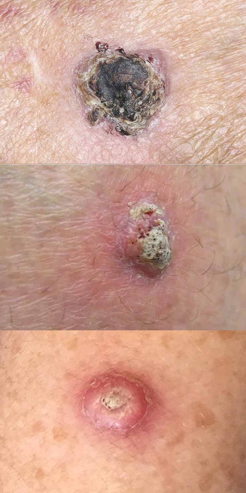
Squamous cell carcinoma varies in appearance. Some lesions resemble a scaly patch that does not resolve, while others develop into thickened, wart-like growths or sores that crust, bleed, or repeatedly reopen. The affected area may be tender or painful, although some lesions cause little discomfort.
Common signs and symptoms include:
- A rough, scaly, or thickened patch of skin that persists or enlarges.
- A firm, raised bump that may have a crusted or depressed center.
- An open sore that does not heal or repeatedly bleeds.
- A wart-like growth that changes in size or appearance.
- A tender or painful lesion, particularly on sun-exposed skin.
- A persistent rough, crusted, or ulcerated area on the lip or ear.
On darker skin tones, SCC may appear brown, violet, red, or skin-colored and may occur in areas with limited sun exposure, including sites of chronic injury or scarring. A suspicious lesion cannot be reliably diagnosed by its appearance alone. Persistent or changing growths deserve appropriate medical evaluation.
What Causes Squamous Cell Carcinoma?
Most cutaneous squamous cell carcinomas develop after accumulated damage to the DNA of skin cells, commonly caused by ultraviolet radiation from sunlight or tanning devices. Over time, these changes can allow abnormal cells to multiply and eventually develop into invasive cancer.
Some SCCs develop from precancerous conditions such as actinic keratosis, which often appears as a rough or scaly patch on sun-damaged skin. Squamous cell carcinoma in situ, also called Bowen disease, describes malignant squamous cells confined to the epidermis without invasion into deeper skin layers.
Not every SCC is caused by sun exposure. Certain cancers develop in chronic wounds, burn scars, areas of persistent inflammation, or in people with reduced immune function. Squamous cell cancers involving mucosal surfaces or internal organs are different clinical diseases and are not necessarily managed using the same guidelines as cutaneous SCC.
Risk Factors for Squamous Cell Carcinoma
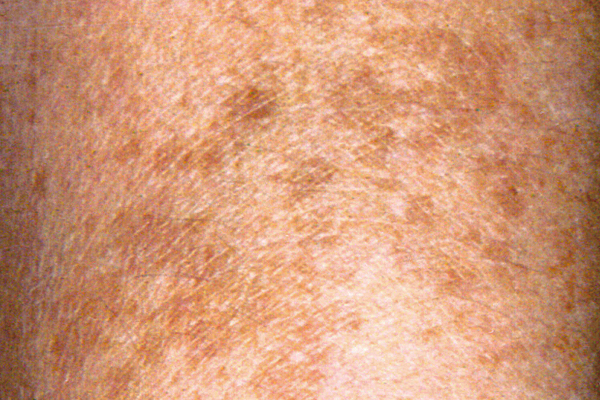
Several factors increase the likelihood of developing cutaneous SCC. Some can be modified through protective habits, while others relate to age, medical history, or inherited susceptibility.
Recognized risk factors include:
- UV Exposure: Years of unprotected sunlight or use of indoor tanning equipment.
- Skin Characteristics: Fair skin, light eyes, blond or red hair, and skin that burns easily.
- Age: Increasing age, reflecting accumulated exposure and other biological changes.
- Previous Skin Cancer: A history of basal cell carcinoma, SCC, or extensive precancerous skin changes.
- Reduced Immune Function: Organ transplantation, certain immunosuppressive medications, and some immune-related medical conditions.
- Chronic Skin Injury: Long-standing wounds, burn scars, and certain inflammatory skin conditions.
- Inherited Conditions: Rare disorders such as xeroderma pigmentosum that increase susceptibility to UV damage.
Common Locations and Higher-Risk Skin Lesions
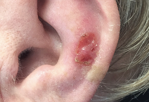
Common locations include the face, ears, lower lip, bald scalp, neck, forearms, backs of the hands, and legs. Some locations, particularly the ear and lip, are associated with greater concern for recurrence or spread. SCC may also develop around nails, on genital skin, or in chronic scars and wounds.
The location of a lesion is only one consideration. A tumor that grows rapidly, becomes large or deeply invasive, shows poorly differentiated cells under the microscope, or grows around nerves may require more extensive treatment and evaluation.
People of every skin tone can develop SCC, and a lack of extensive sun exposure does not eliminate the possibility. Any persistent, enlarging, bleeding, or otherwise suspicious growth should be evaluated by a qualified healthcare professional.
Treatment Options for Squamous Cell Carcinoma of the Skin
Most cutaneous squamous cell carcinomas can be successfully treated when diagnosed early. Treatment depends on the tumor’s size, location, depth, microscopic characteristics, and risk of recurrence or spread, as well as the patient’s health and preferences. Surgery is the principal treatment for most invasive SCCs, while radiation therapy, selected local treatments, and systemic medications may be appropriate in other circumstances.
Surgical Excision and Mohs Surgery
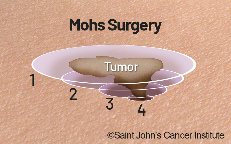
Surgical excision removes the cancer with a margin of surrounding tissue to reduce the likelihood that tumor cells remain. The recommended margin and method of evaluating the removed tissue depend on the tumor’s risk characteristics and anatomical location.
Mohs micrographic surgery removes tissue in stages, with microscopic examination of the surgical margins during the procedure. This technique allows the surgeon to identify remaining cancer while preserving healthy tissue wherever possible. Mohs surgery is often considered for higher-risk SCCs, recurrent tumors, and cancers in areas where tissue preservation and complete margin evaluation are especially important.
For many low-risk SCCs, conventional surgical excision provides effective treatment. The choice of procedure is individualized rather than determined solely by tumor size or location.
Radiation Therapy
Radiation therapy uses high-energy radiation to damage cancer cells. It may be recommended when surgery is not feasible or would produce unacceptable functional or cosmetic consequences, or as additional treatment after surgery for selected higher-risk cases.
Radiation may also help manage cancers that involve regional tissues or lymph nodes. The treatment approach depends on the tumor’s location, extent, pathology findings, and possible effects on nearby healthy structures.
Radiation is not necessary for every SCC and should be considered in the context of the patient’s overall treatment plan.
Other Treatments for Selected Superficial or Low-Risk Lesions
Some less invasive procedures may be appropriate for carefully selected superficial or low-risk skin cancers. These options are not interchangeable with surgery for higher-risk or deeply invasive SCC.
Depending on the diagnosis, selected options may include:
- Curettage and Electrodesiccation: Removal of tumor tissue with a scraping instrument followed by electrical treatment of the affected area, generally reserved for selected small, low-risk lesions.
- Cryosurgery: Destruction of abnormal tissue using extreme cold, considered in limited circumstances for superficial disease or when other approaches are unsuitable.
- Topical Prescription Therapy: Medications such as fluorouracil or imiquimod may be used for certain precancerous lesions or squamous cell carcinoma in situ, but are not established substitutes for surgery in invasive SCC.
- Photodynamic Therapy: A treatment used for selected superficial precancerous lesions or SCC in situ, rather than a routine treatment for invasive disease.
The distinction between SCC in situ and invasive SCC is essential when evaluating these options. Treatment selection should follow a confirmed diagnosis and assessment of the risk of recurrence.
Immunotherapy for Advanced and Selected High-Risk SCC
Immune checkpoint inhibitors have expanded treatment options for patients with cutaneous squamous cell carcinoma that cannot be cured with surgery or radiation, including selected locally advanced and metastatic cases. These medications help the immune system recognize and attack cancer cells.
Systemic immunotherapy options include PD-1 inhibitors such as cemiplimab and pembrolizumab, as well as the PD-L1 inhibitor cosibelimab for eligible patients. The choice of medication depends on the approved indication, disease extent, previous treatment, and individual medical circumstances.
Cemiplimab is also an approved postoperative (adjuvant) treatment for selected adults with cutaneous SCC at high risk of recurrence following surgery and radiation. This does not mean that every patient who undergoes surgery or radiation needs immunotherapy; eligibility depends on specific high-risk features and clinical assessment.
Immunotherapy can produce meaningful responses, but it may also cause immune-related inflammation involving organs such as the lungs, intestines, liver, thyroid, and other endocrine glands. Patients require appropriate evaluation and monitoring.
Other Systemic Treatments for Advanced Disease
For selected patients with locally advanced or metastatic SCC who are not candidates for immunotherapy, or whose disease progresses despite treatment, other systemic options may be considered. These can include certain EGFR-directed therapies or conventional chemotherapy, depending on the circumstances.
These treatments generally have a more limited role than modern immunotherapy for eligible patients. Their potential benefits, risks, and treatment goals should be discussed with an oncology team familiar with advanced cutaneous SCC.
Multidisciplinary Care and Treatment Planning
Patients with complex, recurrent, or advanced squamous cell carcinoma may benefit from coordinated evaluation by specialists in dermatology, surgical oncology, medical oncology, radiation oncology, pathology, and reconstructive surgery. The appropriate treatment may involve more than one approach, particularly when tumors affect important nerves, nearby lymph nodes, or sensitive anatomical structures.
At Saint John’s Cancer Institute, multidisciplinary review can help patients understand their diagnosis, available treatment options, and potential effects on function, appearance, and recovery. Individualized planning helps determine when specialized treatment or a clinical trial may be appropriate.
Reducing Risk and Monitoring Squamous Cell Carcinoma
Many cases of cutaneous squamous cell carcinoma are associated with cumulative UV exposure. While no preventive measure eliminates the risk entirely, consistent skin protection, awareness of suspicious changes, and appropriate dermatologic care can help reduce risk and support early detection. Continued monitoring is particularly important for patients who have already been treated for skin cancer.
Protecting Your Skin From UV Exposure
Sun protection can help reduce additional UV damage to the skin and is especially useful for people who work outdoors, spend substantial time in the sun, or have a history of skin cancer or precancerous lesions.
Recommended protective measures include:
- Seek Shade: Reduce direct sun exposure when UV radiation is strongest, particularly around midday.
- Use Sunscreen: Apply broad-spectrum, water-resistant sunscreen with SPF 30 or higher to exposed skin and reapply approximately every two hours outdoors, or after swimming or sweating.
- Wear Protective Clothing: Use a wide-brimmed hat, UV-protective sunglasses, and tightly woven or UPF-rated clothing.
- Avoid Indoor Tanning: Tanning devices expose the skin to UV radiation that increases skin cancer risk.
- Monitor New or Changing Lesions: Seek evaluation for persistent scaly patches, sores that do not heal, or growths that change or bleed.
For patients with repeated skin cancers or substantial cumulative sun damage, clinicians may discuss additional preventive strategies based on individual risk. Supplements or medications should not be assumed to prevent SCC without considering the evidence and the patient’s medical circumstances.
Skin Examinations and Follow-Up After Treatment

Patients who have had cutaneous SCC are at increased risk of developing additional skin cancers. Follow-up includes examination of the skin and, when clinically appropriate, assessment of nearby lymph nodes or other areas of concern.
The recommended schedule depends on the original tumor’s risk classification, location, treatment, and the patient’s overall medical history. People with higher-risk disease or significant immune suppression may require closer surveillance than those treated for a small, low-risk lesion.
Patients should report new or enlarging lesions, persistent wounds, unusual tenderness, numbness, or swelling near the original tumor site. Your healthcare professional can recommend an examination schedule and explain when additional testing might be appropriate.
Supporting Patients Through Skin Cancer Care
Although most SCCs are treated successfully, the diagnosis may raise concerns about additional procedures, scarring, recurrence, or the possibility of more extensive treatment. Clear explanations of the pathology findings and recommended care can help patients understand their options and prepare for recovery.
For those requiring specialized treatment, Saint John’s Cancer Institute can coordinate evaluation and connect patients with relevant cancer support resources when appropriate.
Get Answers About Changes in Your Skin

A persistent skin lesion, an unexpected biopsy result, or a diagnosis of squamous cell carcinoma can raise questions about whether treatment is needed and what happens next.
At Saint John’s Cancer Institute, our melanoma and skin cancer specialists work together to evaluate complex cases and develop individualized treatment recommendations. Whether you are seeking clarification about a diagnosis, exploring treatment options, or considering a second opinion, our team can help you understand the findings and determine appropriate next steps.
If you have questions regarding squamous cell carcinoma or would like an expert second opinion, please call today or click here to schedule an appointment.