
Understanding Merkel Cell Carcinoma
Merkel cell carcinoma (MCC) is a rare but potentially aggressive form of skin cancer that often develops as a painless, fast-growing bump on the skin. It is also known as cutaneous neuroendocrine carcinoma. Because MCC can spread to nearby lymph nodes or other parts of the body, recognizing suspicious changes and obtaining a timely diagnosis are especially important.
What Does Merkel Cell Carcinoma Look Like?
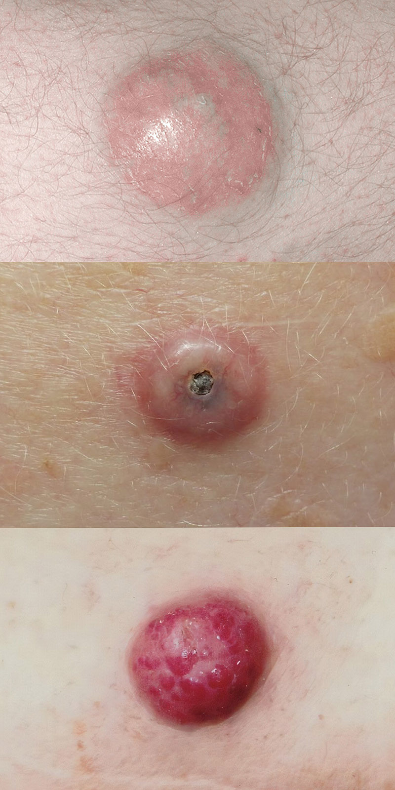
Merkel cell carcinoma commonly appears as a firm, dome-shaped or raised lesion that grows noticeably over weeks or months. The growth may be flesh-colored, pink, red, or violet. Unlike some other skin cancers, it often does not cause pain or discomfort, which can make it easy to mistake for a harmless bump, cyst, or insect bite.
MCC frequently develops on sun-exposed areas such as the head, face, neck, and arms, but it can arise elsewhere on the body. Although it is more common in older adults and people with lighter skin, Merkel cell carcinoma can develop in people of any skin tone.
Changes that deserve medical evaluation include:
- A new, firm skin bump that grows rapidly.
- A painless pink, red, purple, or flesh-colored nodule.
- A growth that resembles a cyst, pimple, or insect bite but persists or enlarges.
- A skin lesion that develops a sore, ulceration, or bleeding.
- A new lump or swelling near the affected area, including nearby lymph nodes.
These findings are not specific to MCC, and many similar lesions are benign. However, a new or rapidly growing skin nodule should be examined promptly rather than monitored indefinitely at home.
What Causes Merkel Cell Carcinoma?
Merkel cell carcinoma develops when changes within skin cells allow abnormal cancer growth. Two major biological pathways are recognized: cancer associated with Merkel cell polyomavirus (MCPyV) and cancer associated with ultraviolet (UV) damage. These pathways help researchers understand how MCC develops, although the precise origin of the cancer cells remains under investigation.
Merkel cell polyomavirus is common and generally harmless. Many people encounter the virus during childhood and never experience symptoms. In rare circumstances, viral genetic material becomes involved in changes that contribute to MCC development. Approximately 70% to 80% of MCC tumors in the United States are associated with this virus. Most remaining cases are linked to extensive UV-related genetic damage.
Having the virus does not mean a person will develop cancer. Merkel cell carcinoma itself is not contagious, and the vast majority of people exposed to MCPyV never develop MCC.
Risk Factors for Merkel Cell Carcinoma
MCC is associated with increasing age, accumulated UV exposure, and reduced immune function. These factors can influence susceptibility, although some patients develop the disease without a clearly identifiable risk factor.
Established risk factors include:
- Advanced Age: Merkel cell carcinoma most commonly affects older adults, particularly those over age 65.
- UV Exposure: Years of exposure to sunlight or artificial ultraviolet radiation can contribute to skin damage and increase risk.
- Weakened Immune Function: People who have received organ transplants, take long-term immunosuppressive medications, or have certain immune-related conditions face increased risk.
- History of Other Skin Cancers: Having previously developed some other skin cancers may be associated with increased MCC risk.
- Skin Characteristics: People with lighter skin that burns easily have a higher observed risk, although MCC can occur in all skin tones.
- Sex: Merkel cell carcinoma is diagnosed more frequently in men than in women.
Unlike many inherited cancer syndromes, most Merkel cell carcinomas are not associated with a recognized hereditary predisposition. Family members do not generally require special screening solely because a relative has received an MCC diagnosis.
For patients with substantial UV exposure or other skin cancer risk factors, routine skin awareness and appropriate dermatologic care are important. A rapidly changing lesion deserves evaluation regardless of whether an individual has known risk factors.
How Merkel Cell Carcinoma Is Diagnosed and Staged
Diagnosing Merkel cell carcinoma requires a biopsy of the suspicious lesion and examination by a pathologist. Because MCC may spread to nearby lymph nodes even when the primary tumor is relatively small, determining the extent of disease is an important part of the initial evaluation. The findings help clinicians recommend treatment and establish an appropriate follow-up plan.
Skin Examination and Biopsy

A healthcare professional examines the skin lesion and nearby lymph nodes, reviews the patient’s medical history, and determines whether a biopsy is necessary. Because MCC can resemble other skin conditions, appearance alone cannot establish the diagnosis.
A skin biopsy removes part or all of a suspicious growth for microscopic examination. A pathologist uses the tissue findings, often including specialized immunohistochemical stains, to distinguish Merkel cell carcinoma from other skin cancers and cancers that may look similar under the microscope.
The pathology report may also describe tumor size, growth characteristics, involvement of surrounding tissues, and whether cancer is present at the edges of the removed specimen. These findings contribute to further evaluation and treatment planning.
Sentinel Lymph Node Biopsy
Sentinel lymph node biopsy (SLNB) is an important staging procedure for many patients with clinically localized Merkel cell carcinoma. It evaluates the first lymph node or nodes that receive drainage from the area of the primary tumor, even when the lymph nodes do not appear enlarged during examination.
During the procedure, a radioactive tracer and, in some cases, a special dye help identify the sentinel lymph nodes. The selected nodes are removed and examined for microscopic cancer cells. Finding MCC in a sentinel lymph node can change the stage and influence treatment recommendations.
Saint John’s Cancer Institute has an established history of research in sentinel lymph node mapping, including techniques developed through the former John Wayne Cancer Institute. This work contributed to broader advances in the surgical staging of cancer. Learn more about Sentinel Lymph Node Biopsy.
Imaging and Additional Testing
Imaging studies may be recommended to evaluate whether MCC has spread to lymph nodes or other parts of the body. Depending on the circumstances, these tests can include PET/CT, contrast-enhanced CT, or other imaging tailored to the patient’s findings.
Additional testing may include blood-based surveillance markers, such as antibodies associated with Merkel cell polyomavirus or circulating tumor DNA (ctDNA), when appropriate and available. These tests may help monitor some patients after treatment, but they do not replace clinical examinations or indicated imaging.
Not every patient requires the same diagnostic procedures. The extent of testing depends on the tumor, physical examination, suspected spread, and available pathology findings.
Understanding the Stages of Merkel Cell Carcinoma
Merkel cell carcinoma is staged according to the primary tumor, involvement of nearby lymph nodes, and whether cancer has spread to distant locations. The staging system helps clinicians estimate risk and select appropriate treatment.
MCC stages are generally described as follows:
- Stage I: A relatively small primary tumor, generally 2 centimeters or less, without identified regional lymph node or distant spread.
- Stage II: A larger primary tumor or one with more extensive local invasion, without identified regional lymph node or distant spread.
- Stage III: Cancer that has involved regional lymph nodes or nearby lymphatic tissue, including certain in-transit tumor deposits, without distant metastasis.
- Stage IV: Merkel cell carcinoma that has spread to distant organs or tissues, such as the lungs, liver, bones, or brain.
These are simplified descriptions. Each stage may have subcategories, and the precise classification depends on clinical examination, pathology, and staging tests. In some cases, MCC may first be identified in a lymph node without a detectable primary skin lesion.
Multidisciplinary Evaluation

Because Merkel cell carcinoma is uncommon and treatment may involve several specialties, patients can benefit from coordinated evaluation. For complex or higher-risk cases, a multidisciplinary team may review pathology, imaging, regional lymph node findings, and potential treatment options.
At Saint John’s Cancer Institute, relevant specialists may include dermatologists, surgical oncologists, medical oncologists, radiation oncologists, pathologists, and reconstructive surgeons. The goal is to develop a care plan appropriate to the patient’s disease characteristics, health, and treatment preferences.
Treatment Options for Merkel Cell Carcinoma
Merkel cell carcinoma treatment depends on the stage of the disease, the location of the tumor, whether nearby lymph nodes are involved, and the patient’s overall health. Treatment may include surgery, radiation therapy, immunotherapy, or a combination of approaches. Because MCC can recur or spread even after an apparently localized diagnosis, treatment planning often includes careful assessment of both the primary tumor and regional lymph nodes.
Surgery and Removal of the Primary Tumor
Surgery is an important treatment for many patients with localized Merkel cell carcinoma. Wide local excision removes the visible cancer along with a margin of surrounding tissue to reduce the likelihood of cancer remaining at the surgical site. The extent of the procedure depends on the tumor’s size, location, and surrounding anatomy.
When sentinel lymph node biopsy is appropriate, it is generally coordinated with the initial surgical treatment so that lymphatic drainage can be accurately evaluated. Patients with cancer in nearby lymph nodes may require additional treatment, such as radiation therapy or selected lymph node procedures.
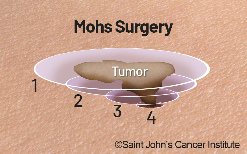
For certain tumors in locations where conserving healthy tissue is especially important, Mohs surgery or other methods of comprehensive peripheral and deep margin assessment may be considered. These techniques examine the surgical margins carefully, but the need for regional lymph node evaluation and appropriate additional treatment remains.
The choice between wide local excision, Mohs surgery, and other local approaches is individualized. MCC care requires attention to the possibility of spread beyond the skin lesion, not simply complete removal of the visible growth.
Radiation Therapy
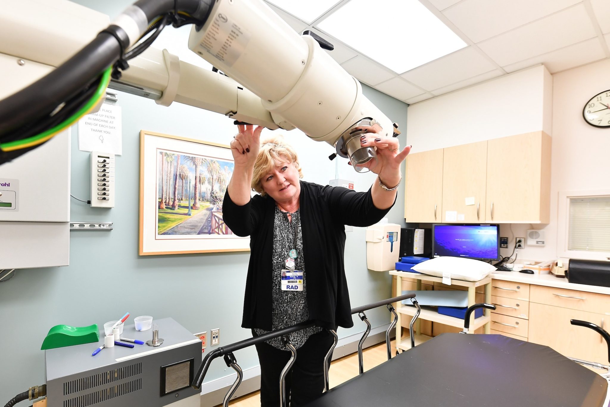
Radiation therapy uses carefully directed high-energy radiation to damage cancer cells. Because MCC can recur near the original tumor site or in regional lymph nodes, radiation may be recommended after surgery for selected patients to reduce the risk of cancer returning in those areas.
Radiation therapy may also be used when surgery is not feasible, when surgical margins cannot be adequately cleared, or to treat selected regional or metastatic sites. The treatment area, dose, and timing depend on the disease extent and potential effects on surrounding tissues.
Some patients with localized MCC may be treated with definitive radiation rather than surgery when the clinical circumstances support that approach. Decisions are best made with input from the relevant surgical and radiation oncology specialists.
Immunotherapy for Advanced Merkel Cell Carcinoma
Immunotherapy has significantly changed the treatment of advanced Merkel cell carcinoma. Immune checkpoint inhibitors help restore the immune system’s ability to recognize and attack cancer cells. For many patients with unresectable locally advanced, recurrent, or metastatic MCC, these medications are preferred over conventional chemotherapy.
Checkpoint inhibitors used in the treatment of advanced MCC include avelumab, pembrolizumab, and retifanlimab. The choice of medication depends on the clinical setting, approval and eligibility criteria, prior treatment, and other medical considerations.
Some patients experience lasting responses to immunotherapy, although not everyone responds. Treatment can also cause immune-related side effects affecting the skin, intestines, lungs, liver, endocrine glands, and other organs. Careful monitoring is necessary throughout treatment.
Clinical trials continue to evaluate immunotherapy before or after surgery for selected patients with earlier-stage or higher-risk disease. These approaches should be discussed in the context of current evidence rather than assumed to be necessary for all patients.
Chemotherapy and Other Treatment Approaches
Chemotherapy has a more limited role in Merkel cell carcinoma than it did before the introduction of immune checkpoint inhibitors. Although MCC can respond to certain chemotherapy regimens, those responses are often less durable, and treatment may cause substantial side effects.
Chemotherapy may still be considered for selected patients who cannot receive immunotherapy or whose cancer progresses despite available treatments. Surgery or radiation may also be used to manage specific tumors or relieve symptoms in patients with advanced disease.
Unlike some other cancers, Merkel cell carcinoma does not currently have a broadly established molecularly targeted treatment that is routinely recommended for most patients. Research continues to investigate additional systemic treatments and combinations.
Clinical Trials and Coordinated Treatment Planning
Because Merkel cell carcinoma is rare, clinical trials are important for evaluating new treatments and determining which approaches offer the greatest benefit for different stages of disease. For eligible patients, participation may provide access to investigational therapies or new ways of combining established treatments.
At Saint John’s Cancer Institute, specialists may coordinate surgical care, radiation therapy, medical oncology, and clinical research opportunities according to each patient’s needs. Patients can review our Clinical Trials information to learn more about research programs and available resources.
Reducing Risk and Monitoring Merkel Cell Carcinoma
Although there is no guaranteed way to prevent Merkel cell carcinoma, protecting the skin from ultraviolet radiation and recognizing new or changing lesions are important steps. For patients who have already received an MCC diagnosis, ongoing follow-up is especially important because the cancer can return locally, involve nearby lymph nodes, or spread to other parts of the body.
Protecting Your Skin From UV Exposure
Ultraviolet radiation is a recognized contributor to Merkel cell carcinoma, particularly in tumors not associated with Merkel cell polyomavirus. Consistent sun protection can reduce UV-related skin damage and support overall skin health.
Recommended protective measures include:
- Seek Shade: Reduce direct sun exposure when UV radiation is strongest, particularly during midday hours.
- Apply Sunscreen: Use broad-spectrum, water-resistant sunscreen with SPF 30 or higher on exposed skin. Reapply approximately every two hours outdoors and after swimming or sweating.
- Wear Protective Clothing: Choose wide-brimmed hats, UV-protective sunglasses, and tightly woven or UPF-rated clothing.
- Avoid Indoor Tanning: Tanning beds and sunlamps expose the skin to harmful ultraviolet radiation.
- Examine Your Skin: Become familiar with existing spots and seek evaluation for any new, persistent, or rapidly changing growth.
Sun protection does not eliminate the risk of MCC, particularly when other biological factors are involved. Nevertheless, it remains an important part of skin cancer risk reduction.
Follow-Up Care After Merkel Cell Carcinoma Treatment
Follow-up after Merkel cell carcinoma treatment is individualized according to the stage at diagnosis, treatment received, immune function, and other risk factors. Regular clinical examinations may include inspection of the skin and assessment of regional lymph nodes. Imaging or blood-based surveillance tests may be recommended for selected patients.
The risk of recurrence is particularly important during the first several years after treatment, although longer-term monitoring may still be necessary. Your care team can establish an examination schedule and explain which symptoms or changes should prompt an earlier visit.
Patients should report new skin nodules, enlarged lymph nodes, unexplained persistent symptoms, or changes near the original tumor site. Prompt evaluation can help determine whether additional testing is necessary.
Supporting Patients Through Treatment and Recovery
A diagnosis of a rare skin cancer can create uncertainty about treatment choices, recurrence, and long-term health. Patients may benefit from clear explanations of their pathology findings, a coordinated treatment plan, and access to supportive services when needed.
At Saint John’s, patients and families can access Cancer Support Services to help address emotional, practical, and physical concerns during cancer care.
Get Answers About Merkel Cell Carcinoma

A new or rapidly growing skin lesion, an unexpected biopsy result, or a confirmed Merkel cell carcinoma diagnosis can raise important questions about what happens next. Understanding the diagnosis, disease stage, and available treatments is the first step toward making informed decisions about your care.
At Saint John’s Cancer Institute, our melanoma and skin cancer specialists work together to evaluate complex cases and develop individualized treatment recommendations. Whether you are seeking an initial evaluation, exploring treatment options, or considering a second opinion, our team can help you understand the findings and determine appropriate next steps.
If you have questions regarding Merkel Cell Carcinoma or would like an expert second opinion, please call today or click here to schedule an appointment.