Minimally Invasive Chest Surgery and Thoracic Surgical Oncology

Welcome to Minimally Invasive Chest Surgery and Thoracic Surgical Oncology. Our surgeons are internationally recognized for lung surgery. With clinical excellence at the heart of our commitment to patient care, our charter encompasses all aspects of thoracic disease, including lung screening and cancer surgery.

CT scan - lung screening - patient education

Our Approach

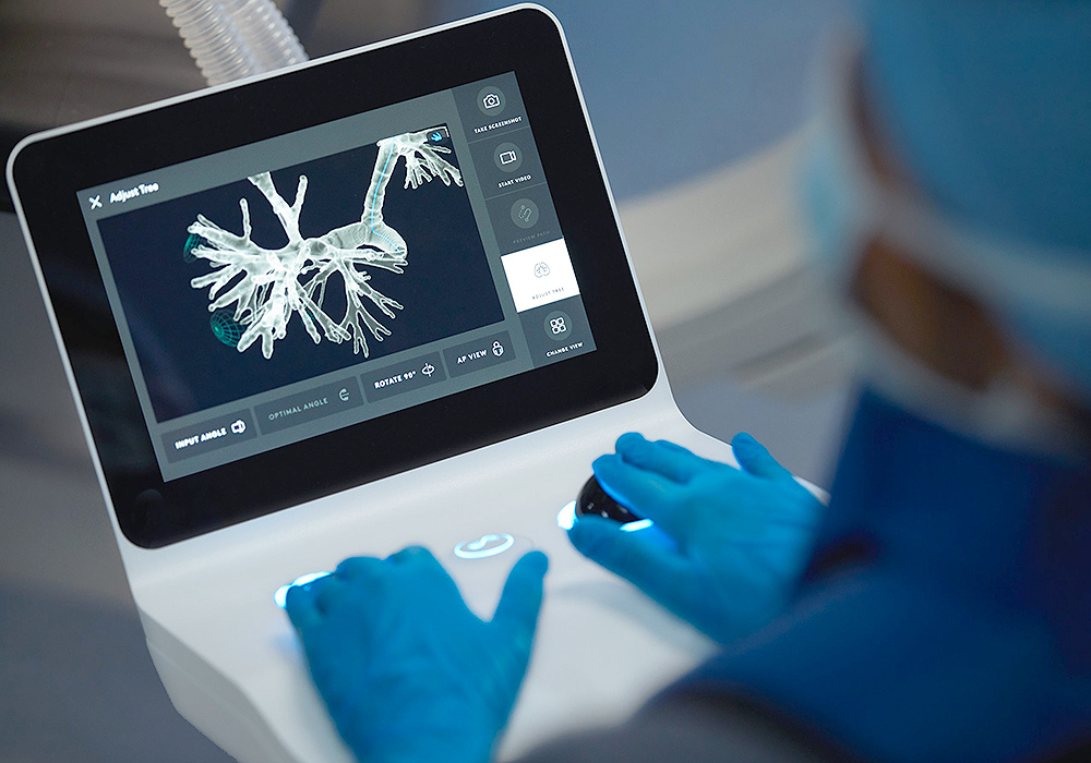
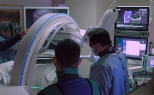
Our commitment is to care for you. As a global leader in thoracic and lung surgery we continue to improve our patient’s quality of life. By utilizing minimally invasive treatment options and advancements in technology, including robot-assisted surgery, transitioning back to normal activities is easier for patients, with shorter recovery time, and fewer side effects.

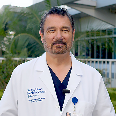
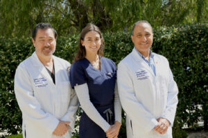
“We are a comprehensive, multidisciplinary Thoracic Center of Excellence that is ready to support you.”

Our thoracic surgical program at Providence, Saint John’s Health Center is “High Performing” in Lung Cancer Surgery, Pulmonology and Lung Surgery, as well as for COPD and Pneumonia,” according to the U.S. News. This distinction is afforded to only those hospitals ranking highest among the Best Regional Hospitals.

With our outcomes-based approach, coupled with our specialized, multi-disciplinary teams and access to innovative resources, such as robot-assisted surgery and translational research, we stress the development of personalized treatment for our patients.

Clinical excellence is our commitment to you.

Surgical Treatments

Minimally Invasive Surgery

Robotic Thoracic Surgery

da Vinci robotic system for lung surgery
The da Vinci robotic surgery system used for lung surgery

Robotic thoracic surgery is robot-assisted, minimally invasive surgery.  This innovation enhances the ability of the surgeon, providing a greater range of motion, surgical precision, and enhanced visualization in less space.  As a result, smaller incisions are required which reduces hospital stay, patient recovery time, and risk of infection. (Learn More)

Lung cancer surgery

Lung cancer surgery may require the removal of a piece of the lung (resection), lobe (lobectomy), or entire lung (pneumonectomy).  These techniques are considered final treatments, which factor in the patient’s total lung function and the amount of lung tissue removal to hinder the spread of cancer.  Lung surgery may also be accompanied by other treatments including radiation therapy and chemotherapy, as with treatment for chest wall tumors. (Learn More) 

Volume Reduction Surgery of the Lung

Patients with severe emphysema can achieve a better quality of life through surgery. Approximately 75 percent of advanced emphysema patients who undergo volume reduction surgery will no longer need an oxygen inhaler after their recovery.  Volume reduction is the resection of both upper lobes of the lungs. (Learn More)

Intuitive Ion System at Providence Saint John’s

What is the Ion system? What does it do?

Intuitive Ion Bronchoscopy - Saint John's Health Center
Intuitive Ion Bronchoscopy is a minimally invasive approach available at Saint John’s Health Center

The Ion system is a robot-assisted surgical tool, that allows surgeons to biopsy or even removes nodules from deep within the lungs, in a less-invasive way than traditional surgical techniques have allowed in the past. A nodule is a group of abnormal cells that have fused together, which may be cancerous.

Also known as the minimally invasive Ion endoluminal system, the Ion is a small, thin, flexible catheter that can be inserted down a bronchoscope while the patient is intubated for surgery. Its tip can move 180 degrees, which allows it to reach all areas of the lungs without the need for additional incisions.

How does it work?

Prior to surgery, the patient has a CT scan, which is used with the Ion’s software program to create an individual “map” of the patient’s lungs. This map is used during surgery to help the surgeon navigate to the nodule deep within the lungs- just like a GPS system for the lungs.

During surgery, the patient is intubated, and a bronchoscope is passed down the throat into the trachea, or windpipe. The Ion catheter is then threaded through the bronchoscope and advanced down into the lungs using a controller. As the Ion catheter is so small and flexible, it can get to areas that were previously very difficult to reach, and which usually needed additional surgical incisions to access.

A camera on the end of the catheter allows the surgeon to visualize the lumens, or airways, of the lungs on a video screen. When the tip of the catheter is advanced to the area where the nodule is located, it is locked into place, and a sample can be taken for biopsy, or the whole nodule can be removed if it is small enough.

At Providence Saint John’s, the Operating Room (OR) has its own pathology department, where testing is done immediately to determine if the nodule is cancerous or not. This real-time confirmation allows our surgeons to make important treatment decisions right in the OR so that the patient does not need to wait and have an additional biopsy or operation.

Why is this good for patients?

As the target nodule can be directly accessed by the Ion through the windpipe and airways of the lungs, one fewer incision is needed to reach it to take a sample for biopsy. This means that less damage is caused to the sensitive lung tissues, which can help to decrease the time needed in-hospital for the patient to recover after surgery.

When lung cancer screening finds a suspicious nodule early on while it is still small, often the whole nodule can be removed using the Ion system, before cancer has a chance to grow and spread. If the nodule is larger or the cancer has spread, the Ion system is used in combination with other minimally invasive surgical techniques, to pinpoint, biopsy, and remove larger areas of tissue that may be cancerous.

Early detection and early surgery can result in a very high five-year survival rate of up to 92% for Stage IA-1 lung cancer, giving the patient a long and healthy life- we can even say the cancer is “cured.”

Surgery for Special Conditions

Benign Esophageal Disease

esophagus conditions and treatments
Conditions of the esophagus may require surgical and non-surgical techniques.

Surgery is not the primary treatment for benign esophageal disease, but more to remedy medical treatments that have not been as effective.  Other procedures involving surgery include vagal-sparing esophagectomy, anti-reflux procedure, and esophageal lengthening.  Primary treatments of benign esophageal disease include medication, dilation (expanding a narrow portion of the esophagus), and the use of stents for conditions where dilation has not been productive.

Bronchiectasis

Bronchiectasis is a chronic respiratory condition characterized by the abnormal widening and scarring of the bronchial tubes, which can lead to the accumulation of mucus, making it difficult to clear the airways and causing recurrent infections. Surgical approaches, including lobectomy, may be necessary for certain cases to prevent further infections and improve overall lung function.

Malignant Esophageal Disease

Surgery to remove cancer of the esophagus may be accompanied by other treatments, such as radiotherapy. Typically, esophageal surgery is common for the removal of very small tumors that have not spread to other areas.  These early-stage cancers can be treated without an external incision using an endoscope.  More developed esophageal cancer may require the removal of a portion of the esophagus (esophagectomy), requiring the stomach to be reconnected to the esophagus.

Myasthenia Gravis

Myasthenia Gravis is a disorder that leads to weakness in muscles, such as in the face, limbs, and eyes.  It can be treated with medication, such as immunosuppressants, and also by removing the thymus gland, which is located in the front-center of the chest.  Surgical removal of the gland (thymectomy) may benefit some individuals by re-balancing the immune system.  Thymectomy is also recommended for individuals with thymoma with mild to moderate muscle weakness.

Mediastinal Tumors

Mediastinal tumors are growths that form in the chest area, surrounded by the breastbone, spine, and lungs on either side. These tumors require surgical resection and may require radiotherapy to help reduce the risk of cancer spread. Minimally invasive surgical techniques, including thoracoscopy or robot-assisted surgery, are common methods of treatment for mediastinal tumors.  Thymic cancers, which forms on the outside surface of the thymus gland, often require surgery, radiation, and chemotherapy.

Thoracic Outlet Syndrome

Thoracic outlet syndrome presents as shoulder and neck pain caused by compressed nerves or blood vessels below the neck.  Typically, non-surgical interventions such as adjusting one’s activities, posture, exercise routine, etc., are recommended.  Surgery is generally a last resort because it involves making structural changes in the body around the area of compression.

Tuberculosis

Tuberculosis (TB) is a highly contagious bacterial infection caused by Mycobacterium tuberculosis. It primarily affects the lungs but can also target other organs and parts of the body. Surgery may be invoved to remove a portion of the affected lung, or the entire lung, in severe cases.

For additional thoracic conditions, please see Thoracic Conditions.


Prevention & Care

New James J. Toth, Sr. Lung Screening Program for Smokers at Saint John’s Health Center

Our new lung screening program is designed to help current and former smokers develop regular examination habits to assess their lung health, including worsening or cancerous conditions. The program also helps non-smokers with screening. Based on your age and smoking habits, screening may be the best way to manage your health moving forward.  Most screening results are negative for cancer, but if lung cancer is found, treatment can begin sooner and may be less invasive, which leads to faster recovery.  Your doctor has greater treatment options and interventions if cancer is discovered early.

Nurse Navigator, Sara Belton, and Dr. Robert McKenna, Director of Minimally Invasive Chest Surgery and Thoracic Surgical Oncology at Saint John’s Health Center, explain the new lung screening program for smokers, highlighting who is most at risk and the benefits of proactive care.

What is the James J. Toth, Sr. Lung Screening Program?

The James J. Toth, Sr. lung screening program at Saint John’s Health Center is for smokers to help identify cancer early, thereby providing greater treatment options and improved outcomes. According to Dr. Robert McKenna, “lung screening has been proven to make a difference.” Dr. McKenna is an internationally renowned thoracic surgeon who has dedicated over 2 decades in General Thoracic surgery research and clinical care. He has produced more than 250 peer-reviewed publications about many different areas of Thoracic Surgery and 2 textbooks in Thoracic Surgery. His book, the Atlas of Minimally Invasive Thoracic Surgery, is a landmark publication in the field of video-assisted thoracic surgery.

Who should be screened for lung cancer?

If you are between 50-75 years old, this age range has the highest risk of developing cancer in the lung. Our program only screens people aged 50-77, with people between the ages of 50 to 60 years being in the most optimal age range for earliest detection. Those eligible for this screening program are current smokers or would have quit smoking within the past 15 years. In addition, the amount of smoking is also important for this screening program, having smoked for at least 20-pack years with no current symptoms of lung cancer.

20-pack years based on smoking one pack of cigarettes per day

20-pack-years-screening-program-infographic-01

If you are a current smoker or quit within the past 15 years, having at least 20-pack years, you are at highest risk.

How do I get screened?

Screening can be arranged with just a phone call. We can help you determine if you qualify as a former or current smoker. Our Nurse Navigator, Sara Belton, Ph.D., will provide additional information and determine if you qualify for the screening program. If you qualify, an appointment will be scheduled for an imaging scan.  One or more physicians will review your scan and will discuss the results of their findings and provide additional information.  Our Nurse Navigator is ready to guide you through the entire process and arrange any follow-up that is needed.

If you have questions regarding lung treatment or our new screening program for smokers, please call today or click here to schedule an appointment

What are the risks & benefits of lung screening for smokers?

Saint Johns Health Center - 80 Years of Healing
Providence, Saint John’s Health Center in Santa Monica, CA – 80 Years of Healing

Lung screening does not prevent cancer but can find early signs of cancer. This allows for earlier treatment interventions of cancer progression, which has been validated to improve patient outcomes. Treatments that may accompany surgery, such as radiotherapy and chemotherapy, can offer greater flexibility for smaller, well-defined tumors, which are more easily managed and even less likely to recur and spread (metastasis). Screening for lung cancer involves using a CT scan which emits a low amount of radiation.

Can you help me quit smoking?

Our Nurse Navigator can connect you with resources to quit smoking. Making a meaningful transition to become a non-smoker is your best first step toward optimal health. Prescriptions for smoking cessation aids can be provided through our thoracic program.

I want to schedule a screening.

Contact our Nurse Navigator at (310) 829-8686 today. A physician’s referral is not required for our lung screening program.