What Are Chest Wall Conditions?
The chest wall is made up of the ribs, sternum, muscles, cartilage, connective tissues, and other structures that protect the lungs and heart and support normal breathing. Chest wall conditions include tumors, infections, structural deformities, and injuries such as rib fractures. These conditions can cause pain, swelling, visible changes in the chest, difficulty with movement, or problems with breathing depending on the structures involved.
Evaluation may include a medical history, physical examination, and imaging such as X-ray, CT, or MRI. Treatment varies widely according to the underlying condition and may range from observation, medication, physical therapy, or antibiotics to surgical repair, tumor removal, or chest wall reconstruction.
Chest Wall Tumors
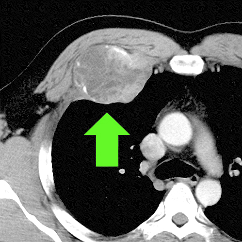
Chest wall tumors are abnormal growths that arise from or involve the bones, cartilage, muscles, connective tissues, or other structures of the chest wall. They may be benign (noncancerous) or malignant (cancerous), and may originate in the chest wall itself or spread there from another part of the body.
Some chest wall tumors cause few symptoms, while others may produce pain, swelling, a palpable mass, or interfere with movement or breathing. Because the type, location, and behavior of these tumors vary considerably, accurate imaging and tissue diagnosis are important before treatment is planned.
Benign and Malignant Chest Wall Tumors
Benign chest wall tumors do not spread to distant parts of the body but may still require treatment if they grow, cause pain, compress nearby structures, or interfere with breathing or movement. Common benign tumors can arise from bone, cartilage, fat, nerves, or other tissues within the chest wall.
Malignant chest wall tumors include several types of sarcoma and other cancers that arise from bone or soft tissue. Sarcomas are among the most common primary malignant tumors of the chest wall. Treatment depends on the specific tumor type, its location, size, and whether it has spread beyond the chest wall. :contentReference[oaicite:1]{index=1}
Primary and Secondary Chest Wall Tumors
Primary chest wall tumors originate in structures such as the ribs, sternum, cartilage, muscle, or connective tissue. Secondary, or metastatic, tumors develop when cancer that began elsewhere in the body spreads to or directly involves the chest wall.
This distinction is important because the treatment of a primary chest wall tumor may differ considerably from treatment of metastatic disease. Evaluation therefore considers both the characteristics of the chest wall mass and the patient’s broader medical and cancer history.
Symptoms of Chest Wall Tumors
Symptoms depend on the tumor’s location, size, rate of growth, and whether nearby structures are involved. Some tumors are discovered incidentally on imaging, while others become noticeable because of a new lump, pain, or changes in the chest wall.
- A new or enlarging chest wall mass
- Localized swelling
- Chest wall pain or tenderness
- Reduced chest movement or discomfort with breathing
- Weakness or loss of muscle function when surrounding tissues are affected
Imaging and Diagnosis
Evaluation generally begins with a physical examination and imaging. Chest X-ray may identify an abnormality involving the ribs or other structures, while CT can provide detailed information about the size and location of a tumor and whether bone or nearby organs are involved.
MRI may be especially useful for evaluating soft-tissue involvement and the relationship of a tumor to muscles, nerves, blood vessels, or other structures. Additional imaging may be recommended depending on the suspected tumor type and whether physicians need to evaluate for disease elsewhere in the body.
Biopsy
A biopsy may be needed to determine exactly what type of tumor is present and whether it is benign or malignant. Depending on the size and location of the mass, tissue may be obtained through an image-guided needle biopsy or a surgical biopsy.
Biopsy planning is particularly important when a malignant tumor such as a sarcoma is suspected, because the location and approach can affect future surgery. For this reason, biopsy is ideally coordinated with the multidisciplinary team that would provide definitive treatment. :contentReference[oaicite:2]{index=2}
Treatment of Chest Wall Tumors
Treatment depends on whether the tumor is benign or malignant, the tissue from which it arises, its size and location, and whether nearby structures are involved. A benign tumor may be observed or surgically removed if it causes pain, grows, interferes with breathing or movement, or affects nearby organs or tissues.
Malignant chest wall tumors often require a multidisciplinary treatment plan that may include surgery, radiation therapy, chemotherapy, immunotherapy, or other systemic treatments depending on the specific cancer. Surgical treatment may involve removing the tumor together with part of the involved rib, sternum, muscle, or surrounding tissue to achieve appropriate margins.
Chest Wall Resection and Reconstruction
When a tumor involves a significant portion of the chest wall, reconstruction may be performed after removal to restore stability, protect the lungs and other organs, and preserve respiratory mechanics. Reconstruction can use nearby muscle or soft tissue, surgical mesh, prosthetic materials, or a combination of techniques depending on the size and location of the defect.
The extent of chest wall removal and reconstruction is individualized and discussed with the patient before surgery. Complex cases may involve collaboration among thoracic surgeons, plastic or reconstructive surgeons, and other specialists. Major thoracic oncology centers use this multidisciplinary approach for extensive chest wall tumors and reconstruction.
For additional information on chest wall tumor treatment, click here.
Chest Wall Infections
Chest wall infections can involve the skin and soft tissues or extend more deeply into structures such as the ribs, sternum, cartilage, and surrounding tissues. Some infections develop after surgery, injury, or another procedure, while others may spread from an infection elsewhere in the chest or body. Because an infection that appears superficial can sometimes involve deeper tissues, persistent pain, swelling, redness, drainage, or fever should be medically evaluated.
Infections involving structures adjacent to the chest wall can also cause similar symptoms. Pleural infection and empyema, for example, occur within the pleural space surrounding the lung and may require treatment by pulmonary and thoracic specialists.
Causes and Risk Factors
Chest wall infections can develop when bacteria, fungi, or other microorganisms enter tissue directly or spread from another site. The source and depth of infection are important because they help determine whether medication alone is appropriate or whether drainage or surgery may also be needed.
Causes and contributing factors may include:
- Previous chest or thoracic surgery
- Trauma or injury to the chest wall
- Infection involving a wound or surgical incision
- Pneumonia or infection within the chest
- Tuberculosis or other specific infections
- Spread of infection from nearby tissues or through the bloodstream
- Conditions or treatments that weaken the immune system
Pleural Infection and Empyema
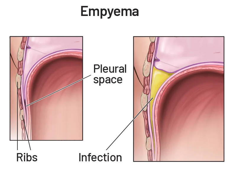
Empyema occurs when infected fluid or pus accumulates in the pleural space between the lung and the inner surface of the chest wall. It most commonly develops as a complication of pneumonia but can also occur following chest surgery, trauma, or other infections.
Treatment depends on the amount and characteristics of the infected fluid and the patient’s overall condition. Antibiotics are generally required, and drainage through a chest tube may be necessary. More complex infections may require additional procedures or thoracic surgery to adequately drain the infection and allow the lung to expand.
Other Causes of Chest Wall and Pleural Pain
Not every painful or inflamed condition involving the chest wall is an infection. Pleuritis, also called pleurisy, is inflammation of the pleura surrounding the lungs and can result from infection as well as other medical conditions. Costochondritis is inflammation of the cartilage connecting the ribs to the sternum and commonly causes localized chest wall pain and tenderness without an underlying infection.
Distinguishing among chest wall infection, pleural disease, musculoskeletal inflammation, and other causes of chest pain is important because their treatments can be very different.
Symptoms of Chest Wall Infections
Symptoms vary according to the location, depth, and severity of the infection. Localized infections may primarily cause pain and swelling, while deeper chest wall or pleural infections can produce more significant respiratory and systemic symptoms.
- Localized chest pain or tenderness
- Swelling, warmth, or redness of the chest wall
- Fever or chills
- Pain that worsens with deep breathing
- Cough
- Shortness of breath
- Drainage from a wound or previous surgical site
Diagnosis of Chest Wall Infections
Evaluation begins with a medical history and physical examination to determine where symptoms originate and whether infection involves superficial tissue, deeper chest wall structures, or the pleural space. Imaging and laboratory testing can then help identify the location and extent of infection and guide treatment.

Diagnostic testing may include:
- Blood tests to look for signs of infection or inflammation.
- Chest X-ray to identify fluid collections, pneumonia, or abnormalities involving the lungs and pleural space.
- CT scan to provide more detailed imaging of the chest wall, lungs, and pleural space and determine the extent of infection.
- Ultrasound to identify and characterize pleural fluid and help guide drainage procedures when needed.
- Fluid or tissue cultures to identify the organism causing an infection and help select appropriate antimicrobial treatment.
- Additional testing when necessary to distinguish infection from cardiac, pulmonary, musculoskeletal, or other causes of chest pain.
Treatment of Chest Wall Infections
Treatment depends on the source, location, and severity of the infection. Antibiotics or other antimicrobial medications may be used when a specific infection is identified or suspected, while pain and inflammation can also be treated as appropriate.
When infected fluid, an abscess, damaged tissue, or a deeper chest wall infection cannot be adequately treated with medication alone, drainage or surgery may be necessary. Thoracic surgery may include removal of infected or damaged tissue, treatment of an infected pleural space, or chest wall reconstruction when infection has significantly affected the ribs, sternum, or surrounding structures.
Chest Wall Structural Conditions
Structural conditions of the chest wall can affect the shape and alignment of the ribs, sternum, and cartilage that form the chest. Some are present from childhood and become more noticeable during growth, while others may develop or become symptomatic later. Depending on their severity, these conditions may primarily affect appearance or may influence posture, exercise tolerance, breathing, and cardiovascular function.
Pectus Excavatum
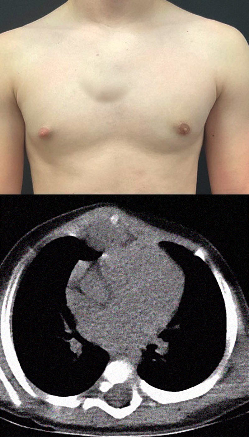
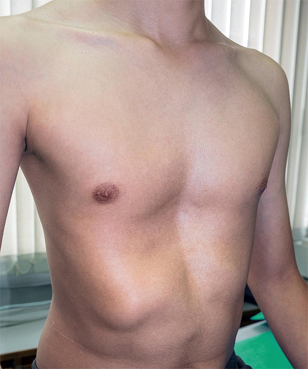
Pectus excavatum is a structural chest wall condition in which the sternum, or breastbone, is displaced inward, producing a sunken appearance of the chest. It is sometimes called funnel chest. The degree of depression can vary considerably and may be relatively symmetrical or more pronounced on one side.
Pectus excavatum is often noticeable during childhood and may become more pronounced during periods of rapid growth. Mild cases may cause few or no physical symptoms, while more significant chest wall depression can reduce the space available for the heart and lungs and contribute to symptoms during physical activity.
Symptoms of Pectus Excavatum
Many people with mild pectus excavatum have no significant physical symptoms. When the chest wall depression is more pronounced, compression or displacement of the heart and reduced space within the chest may contribute to symptoms, particularly during exercise. :contentReference[oaicite:2]{index=2}
- Shortness of breath, particularly with physical activity
- Reduced exercise tolerance or endurance
- Fatigue during activity
- Chest pain or discomfort
- Rapid heartbeat or heart palpitations
- Dizziness or lightheadedness in some patients
Pectus excavatum can also affect body image and self-confidence, particularly during adolescence and young adulthood. These concerns can be an important part of evaluating the overall impact of the condition.
What Causes Pectus Excavatum?
The exact cause of pectus excavatum is not fully understood. Abnormal development of the cartilage connecting the ribs to the sternum is believed to contribute to the inward displacement of the breastbone, and genetics may also play a role because the condition can occur in families.
Pectus excavatum may occur by itself or in association with other skeletal, genetic, or connective-tissue conditions. Recognizing these associations can be important when deciding whether additional evaluation is appropriate.
Associated Conditions
Some people with pectus excavatum have other conditions affecting connective tissue, skeletal development, or the spine. The presence of an associated condition does not necessarily mean that it caused the pectus excavatum, but it may influence the patient’s overall evaluation and care.
- Marfan syndrome – A genetic connective-tissue disorder that can affect the skeleton, heart, blood vessels, and eyes and may be associated with chest wall abnormalities.
- Ehlers-Danlos syndrome – A group of inherited connective-tissue disorders that can affect joints, skin, blood vessels, and other tissues.
- Scoliosis – Abnormal curvature of the spine, which may occur in some people with pectus excavatum.
- Other genetic or skeletal conditions – Certain inherited conditions can be associated with abnormalities of chest wall development.
Diagnosis and Evaluation of Pectus Excavatum
Pectus excavatum can often be recognized during a physical examination, but additional testing may be recommended to determine the severity of the chest wall depression and whether it is affecting the heart, lungs, or exercise capacity. The extent of testing depends on the patient’s age, symptoms, physical findings, and whether treatment is being considered. :contentReference[oaicite:4]{index=4}
- Chest X-ray may provide an initial view of the chest wall and position of the heart.
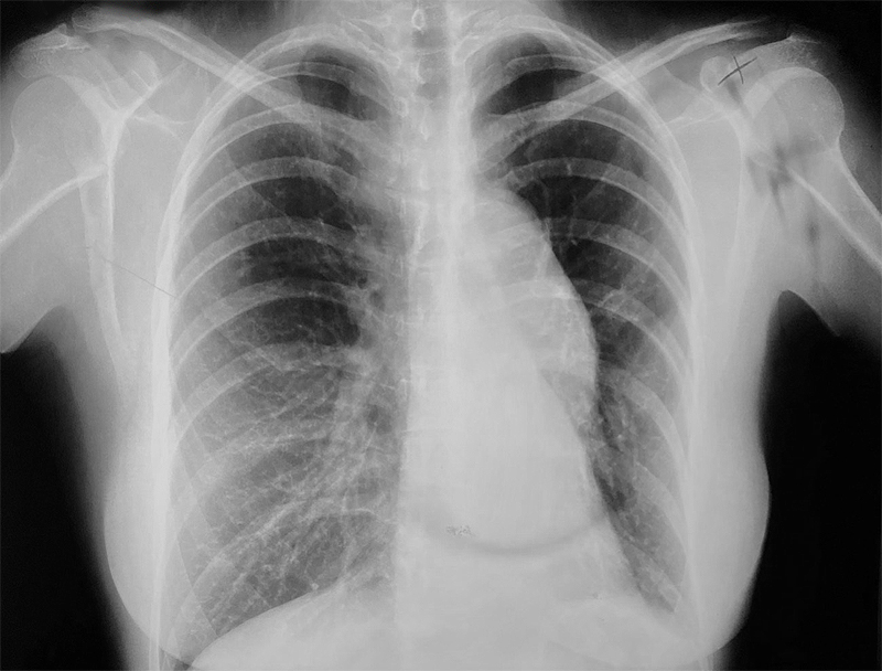
- CT or MRI can show the anatomy of the chest in greater detail, measure the degree of chest wall depression, and evaluate compression or displacement of the heart and lungs.
- Echocardiogram may be used to evaluate heart structure and function and determine whether the chest wall is affecting cardiac function.
- Electrocardiogram (EKG/ECG) may be performed to evaluate heart rhythm and electrical activity when appropriate.
- Pulmonary function tests can evaluate lung volumes and respiratory function.
- Exercise testing may help determine how effectively the heart and lungs function during physical activity.
CT imaging can also be used to calculate measurements such as the Haller index, which helps physicians characterize the severity of chest wall depression. Imaging measurements are considered together with symptoms and functional testing rather than used alone to determine whether treatment is appropriate. :contentReference[oaicite:5]{index=5}
Treatment of Pectus Excavatum
Treatment is individualized according to the severity of the chest wall depression, symptoms, age, heart and lung function, exercise limitations, and the patient’s goals. Mild pectus excavatum may not require surgical treatment, while patients with more significant functional impairment or chest wall compression may benefit from corrective procedures. :contentReference[oaicite:6]{index=6}
Nonsurgical Treatment
Physical therapy may help improve posture, chest mobility, muscle strength, and breathing mechanics, although it does not necessarily correct the underlying structural depression. Selected younger patients with a flexible chest wall may also be candidates for a vacuum bell, a device that applies suction over the sternum to gradually lift the chest wall outward. :contentReference[oaicite:7]{index=7}
Surgery for Pectus Excavatum
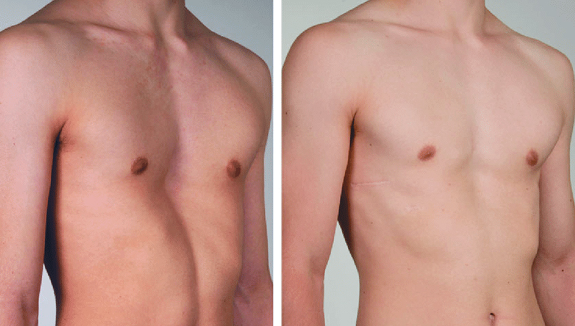
Surgery may be considered when pectus excavatum is moderate to severe, causes significant symptoms or functional limitations, affects the heart or lungs, or has a substantial impact on the patient’s quality of life. The goal is to reposition the sternum, increase space within the chest, and improve chest wall anatomy and function.
Nuss Procedure
The Nuss procedure is a minimally invasive approach performed through small incisions on the sides of the chest. One or more curved metal bars are positioned behind the sternum and used to elevate it into a more normal position. The bars remain temporarily while the chest wall remodels and are removed during a later procedure. :contentReference[oaicite:8]{index=8}
Ravitch Procedure
The Ravitch procedure is an open surgical approach that may be appropriate for selected patients depending on chest wall anatomy, age, previous procedures, or other clinical considerations. The operation involves reshaping or removing abnormal cartilage and repositioning the sternum, which may then be supported with surgical hardware while the chest heals. :contentReference[oaicite:9]{index=9}
Recovery After Pectus Excavatum Surgery
Recovery varies according to the procedure, patient age, and individual anatomy. Pain management, breathing exercises, gradual return to activity, and follow-up with the surgical team are important parts of recovery. Modern pain-management approaches may also be used to improve comfort during the early postoperative period.
As with any chest surgery, potential complications can include bleeding, infection, pneumothorax, pleural effusion, movement or other problems involving implanted hardware, and recurrence of the chest wall depression. The surgical team reviews the expected benefits, recovery process, and individual risks with each patient before treatment.
Rib Fractures and Chest Wall Injuries
Rib fractures are common chest wall injuries that most often occur after a fall, motor vehicle collision, sports injury, or other direct impact to the chest. They can also develop from repetitive stress, severe coughing, or relatively minor trauma in people whose bones have been weakened by osteoporosis or another condition.
Most uncomplicated rib fractures heal without surgery. However, multiple or significantly displaced fractures can affect the stability of the chest wall and make breathing difficult, while the force that caused the fracture may also injure the lungs, blood vessels, or nearby organs. Evaluation therefore considers both the fractured ribs and possible injuries within the chest or upper abdomen.
Symptoms of Rib Fractures
Pain is the most common symptom of a rib fracture and often becomes more noticeable when the chest wall moves. Because significant pain can cause a person to breathe more shallowly or avoid coughing, adequate pain control is important for maintaining normal breathing and reducing the risk of pulmonary complications.
Symptoms may include:
- Localized chest wall pain or tenderness
- Pain with deep breathing or coughing
- Pain when pressing on the injured area
- Pain with bending, twisting, or other movement
- Swelling or bruising over the injured area
- Shortness of breath when the injury is more significant
Causes and Risk Factors
Direct trauma is the most common cause of rib fractures, but bone strength and underlying health can influence how easily a rib breaks. In some patients, a fracture can occur after comparatively minor stress because the rib has already been weakened.
- Falls and accidents – Falls, motor vehicle collisions, and other impacts are common causes of rib fractures.
- Sports injuries – Contact sports and activities that expose the chest to repeated impact or stress can cause rib injuries.
- Osteoporosis – Loss of bone density can make the ribs more susceptible to fracture, sometimes following relatively minor trauma.
- Repetitive stress or severe coughing – Repeated force on a rib can occasionally produce a stress fracture.
- Bone tumors or metastatic cancer – Cancer involving a rib can weaken the bone and increase the risk of a pathologic fracture.
Diagnosis of Rib Fractures
Evaluation begins with a history of the injury and a physical examination of the chest wall and breathing. Imaging may be used to identify fractures and, importantly, evaluate for injuries to the lungs, pleural space, blood vessels, or other structures that may have occurred at the same time.
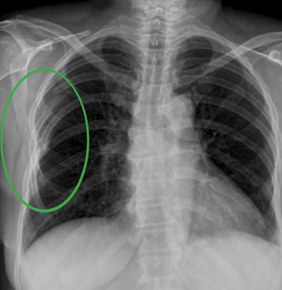
A chest X-ray may identify some rib fractures and complications such as pneumothorax, although small or nondisplaced fractures are not always visible. CT provides more detailed evaluation and can identify fractures and associated chest injuries that may not be apparent on an X-ray. MRI or other imaging may occasionally be used when physicians are evaluating stress fractures, soft-tissue injury, or another underlying condition.
Nonsurgical Treatment
Most rib fractures heal without an operation, often over approximately six to twelve weeks depending on the injury and the patient’s overall health. Treatment focuses on controlling pain while maintaining adequate breathing, coughing, mobility, and activity as healing progresses.
Pain management may combine several approaches to reduce discomfort while limiting unnecessary reliance on opioid medications. Depending on the severity of pain, treatment may include oral medications or regional pain-control techniques such as nerve blocks. Deep breathing, coughing, early mobility, and other pulmonary-hygiene measures are important because shallow breathing caused by pain can increase the risk of atelectasis and pneumonia.
Complications of Rib Fractures
More significant chest injuries can be associated with complications involving the lungs, pleural space, blood vessels, or nearby organs. A displaced rib fragment can occasionally injure surrounding structures, while the original trauma itself may cause pulmonary contusion, pneumothorax, hemothorax, or injury to organs such as the liver or spleen.
Older adults, people with multiple fractures, and patients with underlying lung disease or other medical conditions may have a greater risk of respiratory complications and may require closer monitoring.
Flail Chest
Flail chest is a serious chest wall injury in which multiple rib fractures create an unstable segment of the chest wall. This instability can interfere with normal breathing mechanics and is often associated with significant underlying lung injury.
Patients with flail chest may require hospitalization, intensive pain and respiratory management, and in selected cases surgical stabilization of the fractured ribs.
Surgical Stabilization of Rib Fractures
Only a subset of patients with rib fractures require surgery. Surgical stabilization of rib fractures may be considered for selected patients with flail chest, multiple significantly displaced fractures, chest wall instability, respiratory impairment, persistent severe pain, or other circumstances in which stabilizing the chest wall is expected to improve recovery.
During surgical stabilization, fractured ribs are repositioned and secured using specialized plates and screws to restore chest wall stability and support normal breathing mechanics. The decision to operate considers the fracture pattern, respiratory function, associated injuries, pain, overall health, and response to nonsurgical treatment rather than the number of broken ribs alone.
If you have questions regarding Chest Wall Conditions and Treatment, please call today. Click here to request an appointment.