

Thoracic and Pulmonary Conditions We Treat

Providence Saint John’s Health Center provides comprehensive evaluation and treatment for a wide range of pulmonary and thoracic conditions, from chronic lung and airway disorders to complex chest conditions and thoracic cancers. Our specialists work together to determine the cause of symptoms, establish an accurate diagnosis, and develop an individualized treatment plan.
Care may include advanced imaging and diagnostic procedures, medical management, minimally invasive interventions, thoracic surgery, cancer treatment, or ongoing monitoring depending on the condition. Explore the conditions below to learn more about symptoms, diagnosis, treatment options, and when specialized pulmonary or thoracic care may be appropriate.
Bronchiectasis
Bronchiectasis is a chronic lung condition in which damaged and widened airways make it difficult to clear mucus effectively. This can contribute to persistent cough, increased mucus production, recurrent respiratory infections, and difficulty breathing. Because bronchiectasis can develop from infections, immune disorders, inflammatory diseases, airway obstruction, and other causes, identifying the underlying cause is an important part of care. Treatment can include airway-clearance techniques, medications, treatment of infections and underlying conditions, pulmonary rehabilitation, and surgery in selected cases.
Learn more about Bronchiectasis
Chest Wall Conditions
Chest wall conditions can affect the ribs, breastbone, cartilage, muscles, and other tissues that protect the organs within the chest and support breathing. These conditions include benign and malignant chest wall tumors, infections, structural conditions such as pectus excavatum, and injuries including rib fractures. Symptoms and treatment vary considerably depending on the underlying problem and its effect on breathing, movement, and surrounding structures. Care may range from observation, medication, physical therapy, and pain management to minimally invasive or reconstructive surgery.
Learn more about Chest Wall Conditions
Lung Cancer
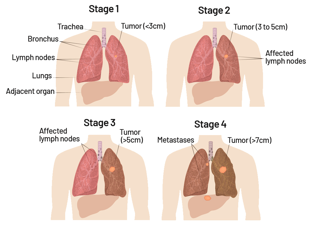
Lung cancer includes several types of cancer that begin in the lungs, most commonly non-small cell lung cancer and small cell lung cancer. Diagnosis may involve advanced imaging, bronchoscopy, robotic or navigational bronchoscopy, endobronchial ultrasound (EBUS), needle biopsy, and genetic and molecular testing to help characterize the cancer. Treatment is individualized according to the cancer type, stage, molecular characteristics, location, and the patient’s overall health and lung function. Options may include surgery, radiation therapy, chemotherapy, immunotherapy, targeted therapy, supportive care, or a combination of treatments.
Lung Cancer Screening
Lung cancer screening can help detect cancer at an earlier stage in people who are at increased risk but do not have symptoms. Low-dose computed tomography (LDCT) is the recommended screening test for eligible individuals with a significant smoking history, although screening recommendations and insurance requirements can differ. Screening also provides an opportunity to discuss smoking cessation, personal risk, and appropriate follow-up if a lung nodule or other abnormality is found. Learn who should consider screening, what to expect during an LDCT scan, and how screening results are evaluated.
Learn more about Lung Cancer Screening
Mediastinal Tumors
Mediastinal tumors and masses develop within the central area of the chest between the lungs and can arise from several different tissues and organs. They include thymic tumors, lymphoma, germ cell tumors, thyroid masses, cysts, neurogenic tumors, and other abnormalities that may be benign or cancerous. The location and appearance of a mass on imaging can provide important clues about its cause and whether a biopsy or additional testing is needed. Treatment depends on the diagnosis and may include monitoring, minimally invasive or robotic surgery, systemic therapy, radiation therapy, or a combination of approaches.
Learn more about Mediastinal Tumors
Paralyzed Diaphragm (Diaphragmatic Paralysis)
Diaphragmatic paralysis occurs when one or both sides of the diaphragm cannot move normally, often because of injury or dysfunction of the phrenic nerve that controls the muscle. Some people have few symptoms, while others experience shortness of breath, difficulty breathing while lying down, fatigue, or reduced exercise tolerance. Evaluation can include pulmonary function testing, chest imaging, diaphragm ultrasound, and a fluoroscopic sniff test to assess diaphragm movement. When symptoms significantly affect breathing and do not improve with conservative care, diaphragm plication may be considered to improve breathing mechanics.
Learn more about Paralyzed Diaphragm
Pulmonary Conditions
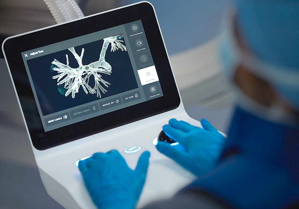
Pulmonary conditions affect the lungs, airways, and the body’s ability to breathe effectively and exchange oxygen. Our pulmonary specialists evaluate and manage conditions including asthma, COPD and emphysema, interstitial lung disease, pulmonary hypertension, airway disorders, pleural disease, pulmonary nodules, respiratory infections, and other complex lung conditions. Diagnosis may involve pulmonary function testing, imaging, bronchoscopy, laboratory testing, and other specialized procedures based on a patient’s symptoms and medical history. Treatment is individualized and may include medication, respiratory therapies, minimally invasive procedures, rehabilitation, surveillance, or coordination with thoracic surgery and other specialists when needed.
Learn more about Pulmonary Conditions
Spontaneous Pneumothorax (Collapsed Lung)
A spontaneous pneumothorax occurs when air enters the space between the lung and chest wall, causing part or all of the lung to collapse without a traumatic chest injury. Symptoms commonly include sudden chest pain and shortness of breath, although severity can vary depending on the size of the pneumothorax and a person’s underlying lung health. Treatment can range from careful observation in selected stable patients to aspiration, catheter or chest-tube drainage, and surgery when air leakage persists or recurrence is a concern. Evaluation can also help identify underlying lung disease or other factors that may increase the likelihood of another pneumothorax.
Learn more about Spontaneous Pneumothorax
Thoracic Outlet Syndrome

Thoracic outlet syndrome (TOS) refers to a group of disorders caused by compression of nerves or blood vessels as they pass between the neck, upper chest, and arm. Depending on whether nerves, veins, or arteries are affected, symptoms can include neck, shoulder, or arm pain, numbness, weakness, swelling, discoloration, or circulation problems. Diagnosis requires determining which structures are being compressed and ruling out other conditions that can cause similar symptoms. Treatment may include physical therapy and activity modification, while vascular complications or persistent compression may require specialized procedures or thoracic outlet decompression surgery.
Learn more about Thoracic Outlet Syndrome
Tuberculosis
Tuberculosis (TB) is an infectious disease caused by bacteria that most commonly affects the lungs but can also involve other parts of the body. Active pulmonary tuberculosis can cause persistent cough, fever, night sweats, fatigue, weight loss, and sometimes coughing up blood, while latent TB infection does not cause symptoms and is not contagious. Treatment primarily involves a carefully selected combination of antimicrobial medications, with the specific regimen depending on whether the infection is latent or active and whether drug resistance is present. Thoracic specialists may become involved when TB causes significant lung damage, complications, or other conditions requiring procedural or surgical evaluation.