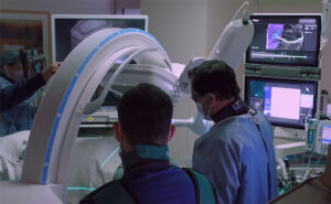

A spontaneous pneumothorax occurs when air enters the space between the lung and chest wall without a traumatic injury, causing the lung to partially or completely collapse. It can occur unexpectedly without known lung disease or as a complication of an existing pulmonary condition.
At Saint John’s Health Center, our pulmonary and thoracic specialists evaluate the extent and possible cause of the pneumothorax, underlying lung health, and risk of recurrence to determine appropriate care. Treatment is based on symptoms, clinical stability, underlying lung disease, persistent air leakage, and previous episodes—not the size of the collapsed lung alone.
What Is a Spontaneous Pneumothorax?
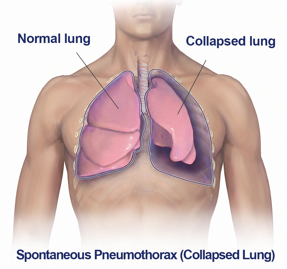
The lungs normally remain expanded against the inside of the chest wall within a thin, fluid-lined area called the pleural space. When air enters this space, pressure can interfere with the lung’s ability to remain fully expanded, resulting in a pneumothorax. Symptoms and severity can vary considerably, from mild chest discomfort and limited shortness of breath to more significant respiratory symptoms requiring immediate treatment.
A spontaneous pneumothorax occurs without an external traumatic injury or medical procedure directly causing the collapse. It may affect only a portion of the lung or involve a more substantial collapse, and the effect on breathing depends on factors such as underlying lung function and whether another pulmonary condition is present.
Types of Spontaneous Pneumothorax
Understanding whether a pneumothorax is primary or secondary can influence both its clinical significance and management. The distinction is based largely on whether established underlying lung disease is present.
Primary Spontaneous Pneumothorax
Primary spontaneous pneumothorax generally occurs in people without known underlying lung disease. Small areas of weakened or abnormal lung tissue, often described as blebs or bullae, may rupture and allow air to escape from the lung into the pleural space.
It occurs more commonly in younger adults and has historically been associated particularly with tall, thin males, although spontaneous pneumothorax can occur in people of different ages and body types. Smoking substantially increases the risk and is an important modifiable factor.
Secondary Spontaneous Pneumothorax
Secondary spontaneous pneumothorax occurs in association with an established lung disorder. Conditions such as chronic obstructive pulmonary disease (COPD), emphysema, cystic lung diseases, pulmonary infections, and other disorders that damage or alter lung tissue can increase susceptibility to pneumothorax.
Because patients with underlying lung disease may already have reduced respiratory reserve, even a pneumothorax that appears relatively limited on imaging can produce significant symptoms. Management therefore considers the patient’s overall pulmonary health and clinical stability rather than simply the apparent size of the collapsed area.
Recurrent Pneumothorax
A person who has experienced one spontaneous pneumothorax has a greater likelihood of experiencing another than someone who has never had one. Recurrence can occur on the same side or, less commonly, affect the opposite lung.
A recurrent pneumothorax may change the discussion about treatment because preventing another episode becomes increasingly important. Previous episodes, persistent air leakage, occupation, lifestyle, and individual risk factors can all influence whether thoracic surgical evaluation is recommended.
Symptoms of a Collapsed Lung
Spontaneous pneumothorax often begins suddenly. Symptoms depend on the amount of lung affected, how quickly air accumulates, and the patient’s underlying respiratory health.

Common symptoms may include:
- Sudden or sharp chest pain, often on the affected side
- Shortness of breath
- Difficulty taking a deep breath
- Chest tightness or discomfort
- Rapid breathing
- Rapid heart rate
- Fatigue or reduced exercise tolerance
- Cough in some patients
Some small pneumothoraces produce relatively mild symptoms, particularly in otherwise healthy individuals. Patients with underlying pulmonary disease may experience considerably greater breathing difficulty.
Sudden severe chest pain, significant or worsening shortness of breath, faintness, bluish discoloration of the lips or skin, or other signs of respiratory distress require immediate medical evaluation.
What Causes Spontaneous Pneumothorax?
Spontaneous pneumothorax can arise through several different mechanisms. Understanding why it occurred is particularly important when a patient has recurrent episodes, underlying lung disease, unusual imaging findings, or a family history suggesting an inherited disorder.
Blebs and Bullae
Blebs and bullae are air-filled spaces that can develop near the surface of the lung. If one ruptures, air may escape into the pleural space and cause the lung to partially or completely collapse.
These abnormalities may be present without previously causing symptoms and can be associated with primary spontaneous pneumothorax. When surgery is required for recurrent pneumothorax or a persistent air leak, abnormal areas of lung may sometimes be identified and treated as part of the procedure.
Underlying Lung Disease
A variety of pulmonary diseases can weaken or alter lung tissue and increase the risk of secondary spontaneous pneumothorax. COPD and emphysema are important examples, but cystic lung diseases, pulmonary infections, interstitial lung disease, and other structural pulmonary disorders may also be associated with pneumothorax.
Identifying an underlying condition can affect immediate treatment as well as longer-term pulmonary care. This is particularly important because patients with compromised lung function may tolerate a pneumothorax differently from otherwise healthy patients.
Genetic and Inherited Conditions
Occasionally, spontaneous or recurrent pneumothorax can be associated with an inherited disorder. One example is Birt-Hogg-Dubé syndrome, a genetic condition associated with characteristic lung cysts and an increased risk of pneumothorax, along with other clinical findings.
Other inherited and connective-tissue disorders may also be associated with pneumothorax. A pattern of recurrent episodes, family history, characteristic lung cysts, or other findings may prompt consideration of additional evaluation or genetic counseling.
Catamenial Pneumothorax
Catamenial pneumothorax is an uncommon form of recurrent spontaneous pneumothorax associated with the menstrual cycle and is often related to thoracic endometriosis. Episodes characteristically occur close to the onset of menstruation and most often involve the right side of the chest.
Because the relationship between chest symptoms and the menstrual cycle may not initially be obvious, recognizing the pattern can be important to diagnosis. Management may require coordination between thoracic specialists and gynecologic care depending on the patient’s circumstances.
Risk Factors for Spontaneous Pneumothorax
Not every person who develops a spontaneous pneumothorax has an identifiable risk factor. However, several characteristics and underlying conditions are associated with an increased likelihood of an initial episode or recurrence.
- Cigarette smoking
- COPD or emphysema
- Other underlying pulmonary diseases
- Blebs, bullae, or cystic changes in the lungs
- Previous spontaneous pneumothorax
- Family history of spontaneous pneumothorax
- Certain genetic or connective-tissue disorders
- Thoracic endometriosis
- Certain age and body-type patterns associated with primary spontaneous pneumothorax

Smoking is particularly important because it is a modifiable risk factor. Patients who smoke are generally encouraged to stop, both for overall pulmonary health and because continued smoking is associated with an increased risk of another pneumothorax.
How Spontaneous Pneumothorax Is Diagnosed
Diagnosis begins with the patient’s symptoms, medical history, and physical examination and is typically confirmed with chest imaging. Evaluation also helps determine the extent of the pneumothorax and whether underlying pulmonary abnormalities may have contributed to its development.
Medical History and Physical Examination
The clinical team will assess the onset and severity of chest pain or shortness of breath, previous pneumothorax episodes, smoking history, underlying pulmonary conditions, recent procedures or injuries, and other factors that may help determine the cause.
Breathing rate, heart rate, oxygen levels, and overall physiologic stability are also important. These findings help physicians determine not only whether a pneumothorax is present but how urgently it may need to be treated.
Chest X-Ray
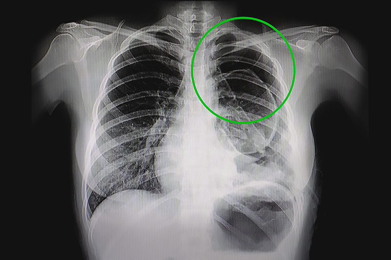
Chest X-ray is commonly used to confirm the presence of a pneumothorax and assess the degree of lung collapse. Imaging may also provide clues about underlying pulmonary disease or other abnormalities.
Follow-up chest imaging may be used to determine whether the pneumothorax is resolving or whether air continues to accumulate. Imaging findings are interpreted together with symptoms and clinical stability rather than used as the sole basis for treatment decisions.
CT and Additional Imaging
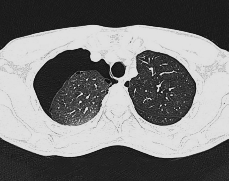
CT imaging provides greater anatomical detail and may be useful when the diagnosis is uncertain, when underlying lung abnormalities are suspected, or when recurrent pneumothorax or surgical treatment is being evaluated.
CT may reveal blebs, bullae, cystic lung disease, or other structural abnormalities that are not as clearly visible on a standard chest X-ray. It is not necessarily required for every uncomplicated spontaneous pneumothorax and is selected according to the individual clinical situation.
Treatment for Spontaneous Pneumothorax
Treatment is individualized according to symptoms, physiologic stability, underlying lung health, whether the pneumothorax is primary or secondary, and whether there is a persistent air leak or history of recurrence. Contemporary management has moved away from treating pneumothorax size as the only deciding factor.
For selected patients with a primary spontaneous pneumothorax who are minimally symptomatic and clinically stable, careful observation may be appropriate even when imaging shows a larger pneumothorax. Other patients may require aspiration, drainage, hospitalization, or surgery, while secondary spontaneous pneumothorax often requires a different level of caution because underlying lung disease may reduce respiratory reserve.
Observation and Conservative Management
Some clinically stable patients with limited symptoms may be managed without immediately removing the air from the pleural space. The body can gradually absorb pleural air while the source of the leak seals, allowing the lung to re-expand over time.
Conservative management requires appropriate patient selection, observation, clear discharge instructions, and follow-up. It should not be interpreted to mean that a collapsed lung can simply be ignored; monitoring is an active part of the treatment strategy.
Ambulatory Management
For selected patients with primary spontaneous pneumothorax, outpatient or ambulatory management may sometimes be considered using appropriate devices and structured follow-up. This approach can allow some patients to avoid an extended hospital stay while the pneumothorax resolves.
Ambulatory management requires appropriate clinical expertise, patient selection, and reliable follow-up and is not suitable for every pneumothorax.
Needle Aspiration or Chest Tube Drainage
When air needs to be actively removed from the pleural space, treatment may involve needle aspiration or placement of a catheter or chest tube. Removing the trapped air reduces pressure around the lung and allows it to re-expand.
The appropriate method depends on the type of pneumothorax, symptoms, clinical stability, underlying lung health, and local expertise. Treatment decisions are individualized rather than determined by one imaging measurement alone.
Persistent Air Leaks
In some patients, air continues to leak from the lung into the pleural space rather than sealing normally. A persistent air leak may prevent complete lung re-expansion or prolong the need for chest drainage.
The next step depends on the patient’s overall condition, underlying lung disease, duration of the leak, and suitability for surgery. Thoracic specialists may consider additional interventions when an air leak does not resolve with initial management.
Surgery to Reduce the Risk of Recurrence
Thoracic surgery may be considered for recurrent pneumothorax, persistent air leakage, certain bilateral presentations, or when preventing another episode is particularly important. Surgical treatment may also be discussed earlier in selected patients based on their individual risks, priorities, occupation, or lifestyle.
Video-assisted thoracic surgery (VATS) allows the chest to be evaluated through small incisions using a camera and specialized instruments. Depending on the underlying problem, surgery may include removal of leaking blebs or bullae and a procedure such as pleurodesis to help the lung adhere to the chest wall and reduce the likelihood of another collapse.
The decision to proceed with surgery is individualized and considers the patient’s overall pulmonary health, recurrence history, and long-term goals.
Recovery and Reducing the Risk of Recurrence
Recovery varies according to the severity of the pneumothorax, the treatment required, underlying pulmonary health, and whether surgery was necessary. Follow-up is important because resolution on imaging, improvement in symptoms, and the risk of another episode all need to be considered before returning to certain activities.
Follow-Up After a Pneumothorax
Patients typically receive follow-up instructions based on how their pneumothorax was managed. Repeat chest imaging may be recommended to confirm that the lung has re-expanded and that pleural air has resolved.

Patients should also understand which symptoms warrant urgent reevaluation. New or recurrent chest pain and shortness of breath after a previous pneumothorax should not be assumed to be harmless.
Smoking and Recurrence Risk
Stopping smoking is one of the most important steps a patient can take to support lung health and reduce the likelihood of another spontaneous pneumothorax. Smoking is strongly associated with primary spontaneous pneumothorax and can also contribute to COPD, emphysema, and structural damage to the lungs.
Smoking cessation may therefore become part of the longer-term care plan rather than being treated as an unrelated lifestyle recommendation.
Air Travel and Scuba Diving
Changes in atmospheric pressure make air travel an important consideration after pneumothorax. Patients should not fly while a pneumothorax remains unresolved and should receive medical guidance about when air travel can safely resume after imaging confirms resolution.
Scuba diving presents additional concerns because substantial pressure changes can have serious consequences if another pneumothorax occurs underwater. Patients with a history of spontaneous pneumothorax should discuss diving specifically with an appropriate physician before considering a return to the activity.
Returning to Normal Activities
Return to work, exercise, and other activities depends on how the pneumothorax was treated and whether symptoms have completely resolved. Patients recovering after surgery may have different restrictions and recovery timelines than those managed conservatively.
Rather than following a universal timetable, activity recommendations should be individualized according to recovery, imaging findings, pulmonary health, and the type of physical activity involved.
Frequently Asked Questions About Spontaneous Pneumothorax
Can a collapsed lung heal without treatment?
Some spontaneous pneumothoraces can resolve without an invasive procedure because the air leak seals and the body gradually absorbs the trapped air. Conservative management may be appropriate for carefully selected patients with primary spontaneous pneumothorax who have minimal symptoms and remain clinically stable.
That does not mean every collapsed lung can safely be left untreated. Medical evaluation is necessary to determine the type of pneumothorax, its effect on breathing, and whether observation or an intervention is appropriate.
Can spontaneous pneumothorax happen again?
Yes. Having experienced a spontaneous pneumothorax increases the possibility of another episode, although recurrence risk differs considerably among patients.
Previous episodes, smoking, underlying lung abnormalities, genetic conditions, and other clinical factors can influence recurrence risk. Recurrent pneumothorax is one reason a patient may be referred for thoracic surgical evaluation.
Why would a healthy person suddenly develop a collapsed lung?
Primary spontaneous pneumothorax can occur in someone without previously recognized lung disease. Small blebs or bullae near the surface of the lung may rupture and allow air to escape into the pleural space, sometimes without any obvious preceding event.
When pneumothorax is recurrent, occurs in several family members, or is associated with unusual lung findings, physicians may investigate whether an underlying genetic or structural condition is contributing.
When is surgery needed for spontaneous pneumothorax?
Surgery may be considered when a pneumothorax recurs, an air leak persists, the lung does not adequately re-expand, or preventing another episode is particularly important. Patient health, underlying pulmonary disease, occupation, lifestyle, and individual priorities can also influence the decision.
Surgical treatment is therefore not determined by one imaging measurement. The thoracic surgeon considers the entire clinical history and the potential benefits of preventing future episodes.
Can I fly after having a pneumothorax?
Air travel should be avoided while a pneumothorax remains present because changes in cabin pressure can cause trapped pleural air to expand. Patients should wait until the pneumothorax has fully resolved and receive guidance from their medical team before flying.
The appropriate timing depends on clinical recovery and confirmation of resolution, so patients planning travel should discuss it during follow-up rather than relying on symptoms alone.

Spontaneous pneumothorax care is part of the broader Pulmonary and Thoracic Center of Excellence at Saint John’s Health Center. Our specialists evaluate both the immediate respiratory problem and the factors that may have contributed to it, including underlying pulmonary disease, structural lung abnormalities, persistent air leaks, and recurrent pneumothorax.
Across the Center, advanced pulmonary diagnostics, imaging, minimally invasive procedures, and robotic-assisted thoracic surgery provide multiple options for evaluating and managing complex chest and lung conditions. Care and long-term lung health are individualized around each patient’s respiratory health and clinical circumstances. Our goal is treating the immediate problem, optimizing your recovery, and recurrence prevention. Please contact us if you have a concern relating to spontaneous pneumothorax.
If you have questions about Spontaneous Pneumothorax (Collapsed Lung), diagnosis, treatment, or of other pulmonary conditions, please call today. Click here to request an appointment.