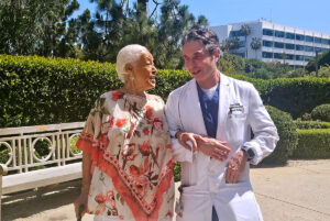

At Providence Saint John’s Health Center, our pulmonary specialists diagnose and treat a broad range of conditions affecting the lungs, airways and breathing. Care ranges from common respiratory diseases such as asthma and chronic obstructive pulmonary disease (COPD) to complex airway disorders, pulmonary nodules, lung infections and cancer.
Our team combines advanced pulmonary diagnostics, interventional pulmonology and coordinated specialty care to determine what is causing a patient’s symptoms and identify the most appropriate treatment.
Pulmonary Conditions We Treat
Some pulmonary conditions affect the airways, while others involve the lung tissue, blood vessels or pleural space surrounding the lungs. Symptoms such as shortness of breath, persistent cough, wheezing or chest discomfort can occur with many different conditions, making an accurate diagnosis important before treatment begins. Our pulmonary specialists evaluate each patient based on their symptoms, medical history, imaging, lung function and other testing as appropriate.
Airway Conditions
Conditions affecting the airways can interfere with the movement of air into and out of the lungs. Saint John’s evaluates and treats airway disorders ranging from common inflammatory conditions to complex narrowing or weakening of the trachea and bronchi.
- Asthma: A chronic inflammatory airway condition that can cause episodes of wheezing, coughing, chest tightness and shortness of breath. Symptoms and severity can vary over time and may be triggered by allergens, respiratory infections, exercise or other factors.
- Bronchial and tracheal stenosis: Narrowing of the trachea or bronchi that can restrict airflow and cause shortness of breath, coughing, wheezing or recurrent respiratory infections. Treatment depends on the location, severity and cause of the narrowing.
- Tracheobronchomalacia: Weakness of the walls supporting the trachea or bronchi that allows the airway to narrow or collapse excessively during breathing. Symptoms may include shortness of breath, persistent cough, wheezing and difficulty clearing mucus.
Chronic Lung Diseases
Chronic lung diseases can affect airflow, damage the airways or reduce the lungs’ ability to function efficiently. Treatment focuses on improving breathing and daily function, reducing symptoms and exacerbations, and slowing disease progression when possible.
- Chronic obstructive pulmonary disease (COPD): A chronic lung disease characterized by persistent airflow limitation, most commonly associated with emphysema and chronic bronchitis. Patients may experience shortness of breath, chronic cough, mucus production and reduced exercise tolerance.
- Emphysema: A form of COPD in which damage to the air sacs of the lungs reduces the surface area available for gas exchange and makes it more difficult to move air out of the lungs. Treatment can include medications, pulmonary rehabilitation and, for carefully selected patients with advanced disease, bronchoscopic or surgical approaches.
- Bronchiectasis: A chronic condition in which damaged and widened airways have difficulty clearing mucus, increasing the risk of recurrent infections and inflammation. Learn more about bronchiectasis.
- Cystic fibrosis: An inherited condition caused by changes in the CFTR gene that can produce thick mucus in the lungs and other organs. Pulmonary care focuses on maintaining airway clearance, treating infections and preserving lung function as part of comprehensive multidisciplinary care.
Interstitial and Pulmonary Vascular Conditions
Some diseases primarily affect the tissue surrounding the air sacs of the lungs or the blood vessels that carry blood through the lungs. These conditions often require specialized testing because shortness of breath and reduced exercise tolerance can have many possible causes.
- Interstitial lung disease (ILD): A broad group of disorders that cause inflammation, scarring or other changes within the tissue of the lungs. The cause and pattern of disease help determine treatment and long-term management.
- Pulmonary hypertension: A group of conditions characterized by elevated blood pressure within the blood vessels of the lungs. Evaluation is important to determine the type and underlying cause because treatment differs considerably among forms of pulmonary hypertension.
Pleural Conditions
The pleura are thin layers of tissue surrounding the lungs and lining the inside of the chest cavity. Disease or inflammation within this space can cause chest pain, shortness of breath or abnormal accumulation of air or fluid.
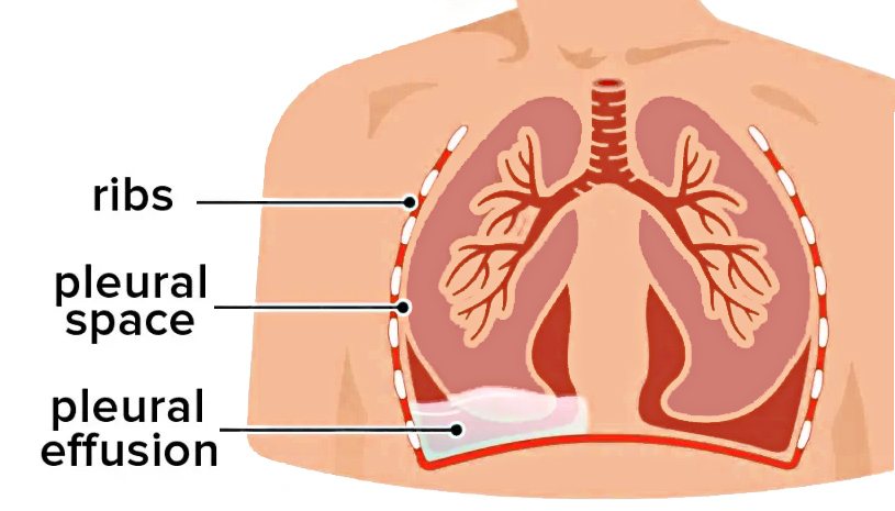
- Pleural disease: Conditions affecting the pleural space include pleural effusion, inflammation and other disorders involving the lining surrounding the lungs. Diagnosis and treatment depend on the underlying cause and whether fluid, infection or another process is present.
- Pneumothorax: Air entering the pleural space can cause part or all of a lung to collapse. Learn more about spontaneous pneumothorax.
Pulmonary Infections
Respiratory infections can range from short-term illnesses to more complex infections requiring specialized pulmonary evaluation. Patients with severe, recurrent or persistent infections may need imaging, laboratory testing or bronchoscopy to identify the cause and guide treatment.
- Pneumonia and other lung infections: Pneumonia occurs when infection causes inflammation within the lungs and may produce cough, fever, shortness of breath and chest discomfort. Treatment depends on the suspected organism, severity of illness and the patient’s overall health.
- Tuberculosis: Tuberculosis is caused by Mycobacterium tuberculosis and most commonly affects the lungs. Evaluation distinguishes inactive TB infection from active TB disease and determines the appropriate treatment. Learn more about tuberculosis.
Pulmonary Nodules and Lung Cancer
Pulmonary nodules are small areas of abnormal tissue commonly discovered on chest imaging. Most nodules are not cancer, but their size, appearance, growth and a patient’s individual risk factors help determine whether they should be monitored or evaluated further. When additional evaluation is needed, advanced imaging, bronchoscopy or biopsy may be used to establish a diagnosis.
- Pulmonary nodules: Nodules may result from prior infection, inflammation, benign growths or lung cancer. Our specialists evaluate suspicious or changing nodules and determine whether imaging surveillance or additional diagnostic testing is appropriate.
- Lung cancer: Saint John’s provides multidisciplinary evaluation and treatment for lung cancer, including advanced diagnostic bronchoscopy, molecular testing, surgery, radiation therapy and systemic treatments based on the type and stage of cancer. Learn more about lung cancer.
Sleep-Related Breathing Disorders
Breathing disorders can also occur during sleep and affect sleep quality, oxygen levels and overall health. Evaluation helps determine the type of breathing disturbance and the most appropriate treatment.
- Sleep apnea: A disorder in which breathing repeatedly stops or becomes reduced during sleep. Obstructive sleep apnea results from narrowing or closure of the upper airway, while central sleep apnea involves disruption of the signals that regulate breathing.
How Pulmonary Conditions Are Diagnosed
Diagnosing a lung or airway condition begins with understanding a patient’s symptoms, medical history and risk factors. Depending on the condition, evaluation may include imaging, pulmonary function testing, laboratory studies and specialized procedures that allow pulmonologists to examine the airways or obtain tissue for analysis. The goal is to identify the cause of a patient’s symptoms while using the least invasive testing appropriate for the clinical situation.
Endobronchial Ultrasound (EBUS)
Endobronchial ultrasound combines bronchoscopy with ultrasound imaging to examine structures next to the airways, including lymph nodes in the chest. During the procedure, physicians can use a small needle passed through the bronchoscope to collect tissue samples. EBUS is commonly used to diagnose and stage lung cancer and can also help evaluate enlarged lymph nodes, infections and other conditions affecting the chest.
Flexible Bronchoscopy
Flexible bronchoscopy uses a thin, flexible instrument equipped with a camera to examine the trachea and airways inside the lungs. It can help physicians identify inflammation, narrowing, bleeding, infection, tumors or other abnormalities and can be used to collect fluid or tissue samples. In selected cases, bronchoscopy can also be used therapeutically to clear secretions or address airway obstruction.
Lung Biopsy
When imaging alone cannot determine the cause of an abnormality, a tissue sample may be needed to establish a diagnosis. Lung tissue can be obtained in several ways, including bronchoscopy, image-guided needle biopsy or minimally invasive surgery. The location and size of the abnormality, the suspected diagnosis and the patient’s overall health help determine the safest and most appropriate approach.
Rigid Bronchoscopy
Rigid bronchoscopy uses a larger, straight bronchoscope to provide direct access to the central airways and is generally performed under general anesthesia. It can be particularly useful when physicians need to remove an airway obstruction, control significant bleeding, obtain tissue or perform other interventional procedures. The technique may also provide access for specialized instruments used to restore or maintain an open airway.
Robotic Navigational Bronchoscopy
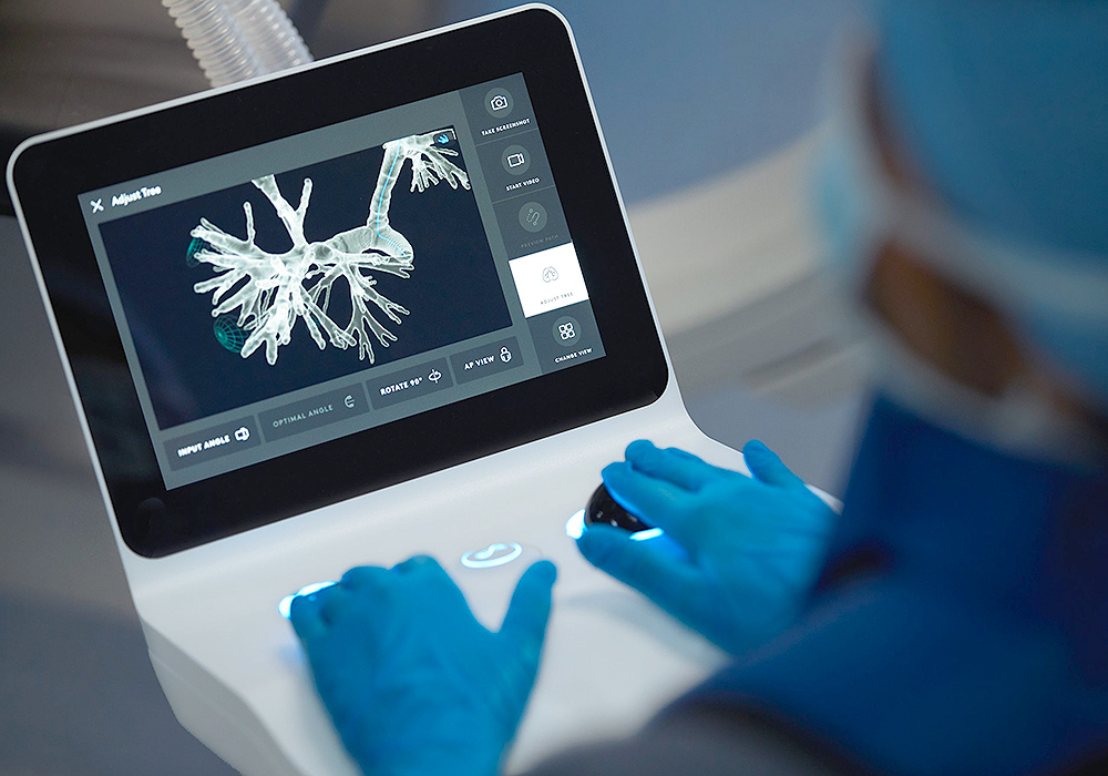
Ion Robotic-assisted bronchoscopy provides advanced navigation and precise control as physicians guide a thin, flexible catheter through branching airways toward suspicious areas within the lung. At Saint John’s, the Ion robotic-assisted bronchoscopy platform can help pulmonologists reach small or difficult-to-access pulmonary nodules and obtain tissue for diagnosis. The procedure is minimally invasive and can play an important role in evaluating abnormalities that may represent lung cancer or other pulmonary disease.
Minimally Invasive Robotic Bronchoscopy and Biopsy
Robotic-assisted bronchoscopy combines advanced navigation, imaging and a highly maneuverable catheter to help physicians reach pulmonary nodules through the natural airways rather than through an incision in the chest. When a suspicious nodule requires further evaluation, the technology can help physicians obtain tissue that can be examined by pathology to determine whether cancer or another condition is present.
Dr. George Chaux discusses how robotic bronchoscopy and advanced imaging help physicians reach and biopsy pulmonary nodules using a minimally invasive approach.
How Are Pulmonary Conditions Treated?
Treatment for pulmonary conditions depends on the diagnosis, severity of disease, symptoms and a patient’s overall health. Care may include medications, respiratory therapies, pulmonary rehabilitation, treatment of an underlying infection or disease, and ongoing monitoring. When a lung or airway condition requires a procedure, our specialists can also use minimally invasive interventional pulmonology techniques to improve breathing, open narrowed airways, drain fluid, obtain or remove abnormal tissue, and treat selected lung conditions.
Interventional Pulmonology and Minimally Invasive Treatment
Interventional pulmonology uses advanced bronchoscopic and image-guided techniques to diagnose and treat conditions involving the lungs, airways and pleural space. Many procedures are performed through the natural airways or through small access points rather than traditional surgery. The appropriate treatment depends on the location and cause of the problem and is selected for each patient individually.
Cryotherapy and Cryoablation
Cryotherapy uses extreme cold to freeze and destroy or remove abnormal tissue. During bronchoscopy, cryotherapy can be used to treat selected airway lesions or remove obstructing tissue. Image-guided cryoablation may also be used in selected patients to treat tumors within the lung, depending on the location of the tumor and the patient’s overall treatment plan.
Laser Therapy
Laser therapy can deliver focused energy through a bronchoscope to remove or reduce abnormal tissue obstructing a central airway. It may be used in selected patients to restore airflow, relieve symptoms or help manage tumors and other lesions involving the trachea or bronchi.
Argon Plasma Coagulation (APC)
Argon plasma coagulation uses ionized argon gas to deliver controlled electrical energy without direct contact with the tissue. During bronchoscopy, APC can be used to control bleeding or treat selected abnormal tissue within the airways and is one of several techniques available for managing central airway obstruction.
Airway Dilation and Stenting
Narrowing of the trachea or bronchi can make breathing difficult and may result from scarring, tumors or other airway disorders. Bronchoscopic dilation can be used to widen a narrowed airway, and in selected cases an airway stent may be placed to help maintain an open passage for airflow. Treatment is individualized based on the cause, location and severity of the obstruction.
Bronchial Thermoplasty
Bronchial thermoplasty is a specialized bronchoscopic procedure that delivers controlled radiofrequency energy to the airway walls. It may be considered for carefully selected adults with severe asthma that remains uncontrolled despite optimized medical treatment. Because treatment options for severe asthma continue to evolve, including advanced biologic therapies, bronchial thermoplasty is considered only after a comprehensive specialist evaluation.
Bronchoscopic Lung Volume Reduction

For carefully selected patients with severe emphysema and lung hyperinflation, bronchoscopic lung volume reduction can provide a minimally invasive alternative to surgery. One-way endobronchial valves are placed in targeted airways to allow trapped air to leave the most diseased portion of the lung while preventing additional air from entering. Reducing hyperinflation can allow healthier areas of the lung and breathing muscles to function more efficiently.
Foreign Body Removal
Objects that are accidentally inhaled can become lodged within the trachea or bronchi and cause airway obstruction, infection or other complications. Flexible or rigid bronchoscopy can be used with specialized instruments to locate and remove the object while minimizing injury to the airway.
Percutaneous Tracheostomy
A tracheostomy creates an opening through the neck into the trachea to provide direct access to the airway. A percutaneous approach can be performed in appropriately selected patients, often in the intensive care setting, when prolonged mechanical ventilation or another condition requires longer-term airway access.
Pleural Catheter
A tunneled pleural catheter is a small, flexible tube placed into the space surrounding the lung to drain recurrent fluid accumulation. It can help relieve shortness of breath caused by pleural effusion and, for selected patients, allows fluid to be managed outside the hospital with appropriate instruction and follow-up.
Speak with a pulmonary specialist. Our team is here to help you understand your condition, explore your treatment options and find the care that is right for you.
If you have questions about pulmonary conditions and diagnosis, please call today. Click here to request an appointment.