

Focused Ultrasound Introduces a New Approach to Treating Liver Tumors
Histotripsy is expanding the options available to selected patients with liver tumors at Providence Saint John’s Health Center. Unlike surgical removal or thermal ablation, this image-guided treatment uses focused ultrasound pulses to mechanically destroy targeted tissue without an incision or insertion of an ablation needle into the tumor. Introduced at Saint John’s in 2025, the technology brought together physicians from several cancer specialties to evaluate its role in treating primary liver tumors and cancers that have spread to the liver from other organs.
In this original physician interview, Jennifer Linehan, MD, explains how histotripsy works, why it offers a different method of tissue destruction, and how researchers are investigating potential effects on the body’s immune response. The article also recognizes the support of Saint John’s Health Center Foundation and the Riordan Foundation in bringing the technology to the Santa Monica community.
What Is Histotripsy?
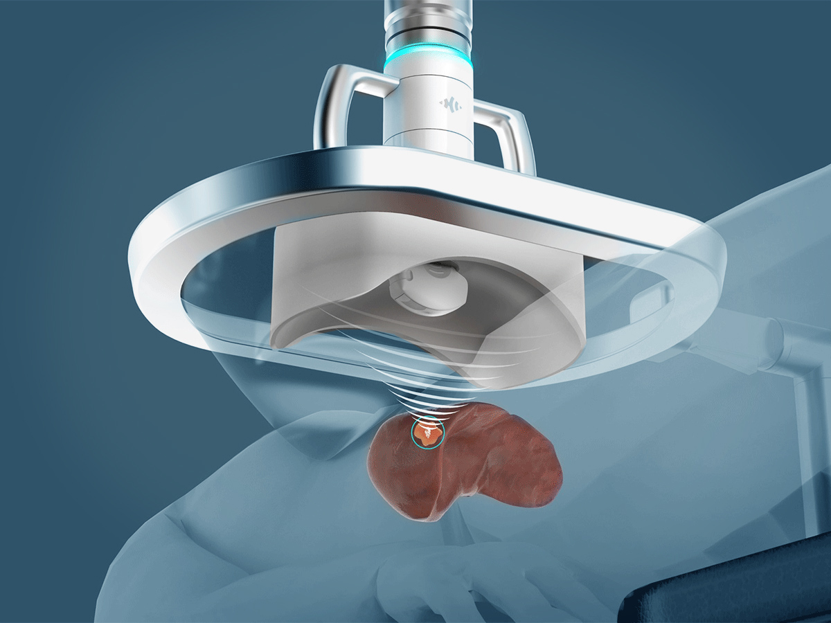
Histotripsy is a noninvasive, nonthermal form of focused ultrasound therapy that uses carefully controlled sound waves to disrupt targeted tissue.
The Edison Histotripsy System, developed by HistoSonics, received FDA authorization in October 2023 for the noninvasive destruction of liver tumors, including tumors that cannot be surgically removed.
This authorization applies to the device’s ability to destroy targeted liver tissue. It should not be interpreted as a guarantee that the treatment cures liver cancer or prevents recurrence.
Histotripsy differs from thermal ablation, which destroys tissue by heating or freezing it. Instead, the system generates rapid changes in pressure that produce and collapse microscopic gas bubbles within the selected treatment area.
How Ultrasound Creates Cavitation Bubbles

During histotripsy, short pulses of high-intensity ultrasound energy are focused within a defined volume of tissue.
The changing pressure creates microscopic bubbles that rapidly expand and collapse. These forces break apart the cellular structure of the targeted tissue without relying on heat as the primary mechanism of destruction.
The physician uses imaging to plan treatment boundaries and monitor the procedure. This helps focus treatment on the intended area while accounting for nearby liver tissue and other anatomical structures.
Although histotripsy can provide precise tissue targeting, treatment is not appropriate for every location. Ultrasound visibility, tumor size, nearby organs, and the ability to reach the lesion with a suitable acoustic pathway all influence patient selection.
A Procedure Without Surgical Incisions
The treatment system delivers ultrasound energy from outside the body. A water-based coupling system helps transmit sound waves toward the targeted tissue within the liver.
General anesthesia is commonly used so the patient remains comfortable and still while the physician controls targeting and treatment delivery.
Procedure time varies according to tumor size, location, the planned treatment volume, and the number of lesions requiring treatment.
Because tissue destruction occurs without an external incision or needle entering the tumor, many patients may experience a shorter recovery than they would after major liver surgery. However, histotripsy remains a medical intervention requiring anesthesia, appropriate monitoring, and evaluation for possible complications.
Dr. Jennifer Linehan Explains the Technology
Urologic oncologist Jennifer Linehan, MD, describes the unusual nature of the procedure in the original interview:
“I tell them this is “Star Wars” technology because there are no cuts, there’s no poking, there there’s nothing that’s going inside you from outside. It’s all happening with a focused beam of ultrasound to destroy the tumors.”
– Jennifer Linehan, MD
Her description emphasizes the defining feature of histotripsy: ultrasound energy is delivered from outside the body rather than through a surgical incision or probe inserted directly into the tumor.
Despite this advantage, the procedure still requires careful patient selection and should not be considered free of medical risks.
Who May Be a Candidate for Liver Histotripsy?
Histotripsy may be considered for selected patients with primary liver cancer or metastatic tumors involving the liver.
A primary liver cancer begins within liver tissue. A liver metastasis occurs when cancer originating elsewhere in the body spreads to the liver.
Examples of cancers that may produce liver metastases include colorectal, breast, pancreatic, kidney, and other malignancies.
Histotripsy treats the targeted tissue within the liver. It does not automatically treat cancer elsewhere in the body, and its role must be evaluated within the patient’s overall cancer care plan.
Primary and Metastatic Liver Tumors
Published clinical studies have included patients with hepatocellular carcinoma, a primary liver cancer, as well as patients with tumors that spread to the liver from other organs.
The presence of a liver tumor alone does not establish eligibility. The care team must determine whether the lesion can be safely visualized and targeted and whether localized treatment is appropriate for the patient’s disease.
For patients with advanced cancers involving multiple organs, histotripsy may be considered alongside systemic therapy rather than as a replacement for medications that treat cancer throughout the body.
How Physicians Determine Whether Histotripsy Is Appropriate
Assessment typically considers:
- Tumor location: Whether the lesion can be adequately visualized and reached with focused ultrasound.
- Tumor size and number: The amount of tissue requiring treatment and whether multiple lesions can be managed appropriately.
- Surrounding structures: The relationship to blood vessels, bile ducts, bowel, ribs, and other nearby anatomy.
- Liver function: The condition of the surrounding liver and the patient’s ability to tolerate treatment.
- Overall cancer status: Whether local treatment fits the management of any disease outside the liver.
- Other treatment options: The expected benefits and risks of surgery, thermal ablation, embolization, radiation, or systemic therapy.
Histotripsy may be especially worth discussing when conventional surgery or another local treatment carries substantial risk, although it is not automatically the preferred option in those circumstances.
Multidisciplinary Treatment Planning
At Saint John’s, evaluation of liver tumors can involve surgical oncology, interventional radiology, medical oncology, radiation oncology, and other specialists according to the patient’s diagnosis.
Collaboration helps determine whether histotripsy should be offered, how it fits with other treatments, and what imaging or additional therapy may be necessary afterward.
Learn more about Liver Cancer and the Institute’s Liver Cancer Treatment Options.
Watch Dr. Linehan Explain How Histotripsy Treats Liver Tumors
In this original 2025 physician interview, Dr. Linehan discusses the focused ultrasound technology, its potential benefits for selected patients, and the research exploring whether mechanical tumor destruction can influence immune activity.
The discussion reflects the introduction of the histotripsy program at Providence Saint John’s Health Center.
How Histotripsy Uses Focused Ultrasound to Destroy Targeted Tumors
Jennifer Linehan, MD, explains histotripsy for selected liver tumors and the potential applications being investigated in cancer research. Original physician interview, 2025.
Could Histotripsy Become a Treatment for Kidney Cancer?
Although histotripsy was introduced at Providence Saint John’s Health Center for the treatment of selected liver tumors, researchers are also studying its potential application in kidney cancer. This emerging area of investigation is particularly relevant to urologic oncology, where treating small renal tumors while preserving healthy kidney tissue is an important clinical objective.
Jennifer Linehan, MD, a urologic surgical oncologist at Saint John’s Cancer Institute, has experience treating kidney cancer through robotic and kidney-preserving surgical approaches. The development of histotripsy for renal tumors represents a potential future extension of focused ultrasound technology into her specialty.
Clinical Trials Are Evaluating Histotripsy for Kidney Tumors
Two clinical studies, CAIN (NCT05432232) and HOPE4KIDNEY (NCT05820087), have investigated the use of histotripsy to treat selected small, nonmetastatic kidney tumors measuring 3 centimeters or less.
The CAIN feasibility study reported encouraging preliminary results involving the ability to target kidney tumors using focused ultrasound. HOPE4KIDNEY is a larger multicenter trial evaluating treatment effectiveness and safety, with longer-term follow-up planned.
These studies will help determine whether histotripsy can safely and effectively destroy kidney tumor tissue while preserving surrounding structures and renal function.
Potential Advantages and Important Limitations
Because histotripsy does not rely on thermal energy, researchers are investigating whether it could offer advantages in selected kidney tumor locations. Its focused, noninvasive treatment method may also provide opportunities for preserving healthy kidney tissue.
However, important questions remain about treatment safety, kidney function, long-term cancer control, tumor accessibility, and how histotripsy compares with established approaches such as partial nephrectomy, cryoablation, and radiofrequency ablation.
As of November 2026, histotripsy for kidney tumors remains investigational in the United States and is not an established alternative to standard kidney cancer treatment.
Learn more about Kidney Cancer Diagnosis and Treatment at Saint John’s Cancer Institute.
Can Histotripsy Influence the Immune System?
One area of ongoing research concerns whether the mechanical disruption of cancer tissue can stimulate immune activity beyond the directly treated region.
Histotripsy breaks apart tumor cells and their internal structures. Researchers are studying whether the resulting biological material and inflammatory signals may help the immune system recognize cancer cells.
This possibility has attracted interest in combining histotripsy with immunotherapy or other systemic treatments.
What Is the Abscopal Effect?
The abscopal effect describes a phenomenon in which treatment directed at one tumor is followed by regression of cancer at another site that was not directly treated.
It has historically been discussed in connection with radiation therapy, particularly when immune activation may contribute to effects beyond the irradiated area.
Researchers are investigating whether nonthermal tumor destruction through histotripsy could produce related immune responses.
Preclinical studies and early clinical observations have provided reasons to study this possibility, but a predictable, clinically established abscopal effect from histotripsy has not been demonstrated in humans.
Dr. Linehan Discusses a Potential Research Benefit
In the original interview, Dr. Linehan describes the scientific question being explored:
“One of the unique things about histotripsy, we suspect, that also does happen with radiation, is it creates what we call an abscopal effect, where because it causes inflammation in that area where you’ve treated the tumor it makes we believe immunotherapies some sometimes even chemotherapies work better.”
– Jennifer Linehan, MD
Dr. Linehan presents this as a suspected biological effect. The research question remains relevant, but patients should not expect histotripsy to reliably shrink untreated tumors or improve immunotherapy outcomes.
The direct purpose of histotripsy is destruction of the selected treatment volume. Effects on distant tumors, survival, and combinations with systemic therapies require further investigation.
Why This Research Matters
Local tumor treatment and systemic cancer treatment have different roles.
Histotripsy is intended to mechanically disrupt selected tissue within the liver. Immunotherapy, chemotherapy, and targeted medications may treat cancer cells throughout the body according to the diagnosis and chosen regimen.
If future research demonstrates that histotripsy reliably enhances systemic immune responses, it could influence how local and systemic treatments are combined.
For now, treatment decisions should be based on established clinical evidence and the patient’s medical needs rather than an anticipated abscopal effect.
What Do Clinical Studies Show About Histotripsy?
Clinical research supports histotripsy’s ability to destroy selected liver tumor tissue, but evidence on longer-term cancer outcomes continues to develop.
The HOPE4LIVER study was an important prospective multicenter trial evaluating the treatment of primary and metastatic liver tumors.
Early Technical Success and Safety Results
In the initial HOPE4LIVER pivotal analysis, researchers reported technical success in approximately 95% of the evaluated tumors.
Technical success meant that the planned treatment area had been adequately covered by histotripsy according to the study’s immediate imaging assessment.
Procedure-related major complications occurred in approximately 7% of the treated patients.
These findings supported early clinical adoption, but technical success does not mean that every patient is cured or that the cancer cannot recur.
One-Year Local Tumor Control
A subsequent HOPE4LIVER analysis reported one-year local tumor control of approximately 90% using a post hoc imaging assessment. A prespecified primary assessment produced a lower estimate of approximately 63%.
The difference illustrates the challenges involved in interpreting images after a relatively new form of tissue ablation.
These findings are encouraging but should be interpreted in the context of a small, selected study population, differences in analysis methods, and limited longer-term follow-up.
What Histotripsy Studies Do Not Yet Establish
Existing evidence does not establish that histotripsy is superior to liver surgery, thermal ablation, embolization, or other liver-directed treatments for all patients.
It also does not demonstrate that every tumor is suitable for treatment, that one session will always be sufficient, or that the procedure reliably produces a systemic immune response.
Continued research is evaluating durability of local control, patient selection, repeat treatment, and the role of histotripsy alongside other cancer therapies.
Potential Benefits, Side Effects, and Recovery
Histotripsy offers a different way of treating selected liver lesions because it does not require an external surgical incision, insertion of an ablation needle into the tumor, or thermal destruction of the target tissue.
Some patients may return home the same day and resume activities relatively quickly, depending on their medical condition and the treatment performed.
However, noninvasive delivery does not mean the procedure is free of side effects or complications.
Potential Benefits
- No external surgical incision or needle placement into the tumor.
- Nonthermal mechanical destruction of targeted tissue.
- Real-time ultrasound visualization during treatment planning and delivery.
- Potential preservation of more surrounding healthy liver tissue in appropriately selected cases.
- Possible same-day discharge and shorter recovery than major surgery for some patients.
- The ability to integrate selected liver-directed treatment with broader cancer care.
Potential Complications
Reported or potential complications can include:
- Pain or discomfort after treatment.
- Temporary changes in liver function or liver enzyme measurements.
- Fever or inflammatory symptoms.
- Bleeding or vascular complications.
- Infection.
- Injury to nearby organs or anatomical structures.
- Complications associated with anesthesia or underlying medical conditions.
Serious complications were uncommon in early published clinical studies but did occur. Risks depend on the tumor’s location, the treatment volume, and individual medical factors.
Follow-Up After Histotripsy
After treatment, physicians generally use follow-up imaging to evaluate the treated area, identify possible residual viable tumor, and monitor for recurrence or new lesions.
Patients with metastatic cancer may also continue systemic treatment and monitoring of disease outside the liver.
The follow-up plan should be tailored to the original cancer diagnosis, treatment intent, and overall condition.
For details about treatment planning and recovery, visit Histotripsy for Liver Tumors.
The Multidisciplinary Histotripsy Program at Saint John’s
The introduction of histotripsy at Providence Saint John’s Health Center brought together specialists with complementary experience in image-guided procedures, liver surgery, and cancer care.
David Tran, MD — Interventional Radiology

David Tran, MD, is an interventional radiologist whose clinical work includes image-guided cancer therapies, liver-directed Y-90 radioembolization, and tumor ablation.
Interventional radiology plays an important role in evaluating liver tumor accessibility, imaging findings, and procedural treatment options.
Jennifer Linehan, MD — Urologic Oncology

Jennifer Linehan, MD, is a urologic surgical oncologist whose involvement in the original program introduction reflects the interdisciplinary interest in focused ultrasound technology and its future cancer applications.
Although histotripsy for liver tumors belongs within the Gastrointestinal and Hepatobiliary program, Dr. Linehan’s interview also highlights the potential for investigation of this technology in other disease settings.
Anton Bilchik, MD — Gastrointestinal and Hepatobiliary Surgical Oncology

Anton Bilchik, MD, is a surgical oncologist whose work includes cancers of the gastrointestinal tract, liver, and pancreas.
Hepatobiliary surgical expertise contributes to decisions about whether liver tumors are better managed with surgery, histotripsy, another local treatment, systemic therapy, or a combination of approaches.
These physicians were identified in the original 2025 program introduction. Current procedural responsibilities and appointment availability should be confirmed through the relevant clinical departments.
Histotripsy at Saint John’s: From Program Introduction to Patient Treatment
The original March 2025 article introduced the technology and explained the scientific principles behind the new service.
Saint John’s subsequently reported that its first histotripsy patient underwent treatment on April 15, 2025, with Dr. David Tran and the multidisciplinary clinical team.
That milestone marked the transition from program development to patient treatment at the health center.
Read Saint John’s Treats First Patient With Histotripsy for Liver Tumors for the original program milestone.
Recognizing the Donors Who Helped Introduce Histotripsy
Bringing new clinical technology into a health system involves more than purchasing equipment. It also requires physician training, operational planning, multidisciplinary collaboration, and resources to support implementation.
The original Saint John’s histotripsy introduction recognized the generosity of donors who helped make this program possible.
The Riordan Foundation was among the supporters acknowledged for its contribution to the introduction of the histotripsy system at Providence Saint John’s Health Center.
“The Riordan Foundation is proud to support the introduction of the histotripsy system to Saint John’s, empowering the incredible physicians at the health center to continue providing exceptional care for all.”
– Elizabeth Gregory Riordan
The quotation is preserved from the original program announcement and recognizes the Foundation’s support for expanding treatment capabilities at Saint John’s.
Saint John’s Health Center Foundation

Saint John’s Health Center Foundation connects the health center with individuals, families, and community partners who support clinical programs, research, equipment, and patient services.
Its role in the histotripsy initiative reflects how philanthropic support can help bring specialized treatment technologies into the local healthcare setting.
To learn more about the Foundation’s work, visit SaintJohnsFoundation.org or explore Ways to Support Saint John’s.
Expanding Liver Tumor Treatment Through Focused Ultrasound
The introduction of histotripsy in 2025 expanded the liver-directed treatment options available at Providence Saint John’s Health Center. By using focused ultrasound pulses to mechanically destroy targeted tumors, the technology offers a noninvasive alternative for selected patients who may benefit from local treatment. Its potential role in stimulating immune activity remains an important area of research, while published studies continue to clarify treatment durability, safety, and appropriate patient selection.
To learn whether histotripsy may be appropriate for a primary liver tumor or cancer that has spread to the liver, explore Histotripsy for Liver Tumors and the Liver Cancer Treatment Program. Patients seeking an evaluation or second opinion can contact the Institute through its online contact form.
Sources & Further Reading
- U.S. Food and Drug Administration — Edison Histotripsy System, Indications for Use
- Mendiratta-Lala M, et al. HOPE4LIVER Pivotal Trial: Histotripsy of Primary and Metastatic Liver Tumors
- HOPE4LIVER — One-Year Clinical Outcomes
- Histotripsy for Liver Tumors — Systematic Review and Meta-Analysis of Clinical Evidence (2026)
- Saint John’s Cancer Institute — Histotripsy for Liver Tumors
- Saint John’s Cancer Institute — Liver Cancer Treatment
- Saint John’s Cancer Institute — First Histotripsy Patient Treated
- Histotripsy for Renal Cell Carcinoma: From Animal Studies to Clinical Application (2026)
- ClinicalTrials.gov — CAIN: Histotripsy for Small Renal Tumors
- ClinicalTrials.gov — HOPE4KIDNEY: Histotripsy for Primary Renal Tumors