
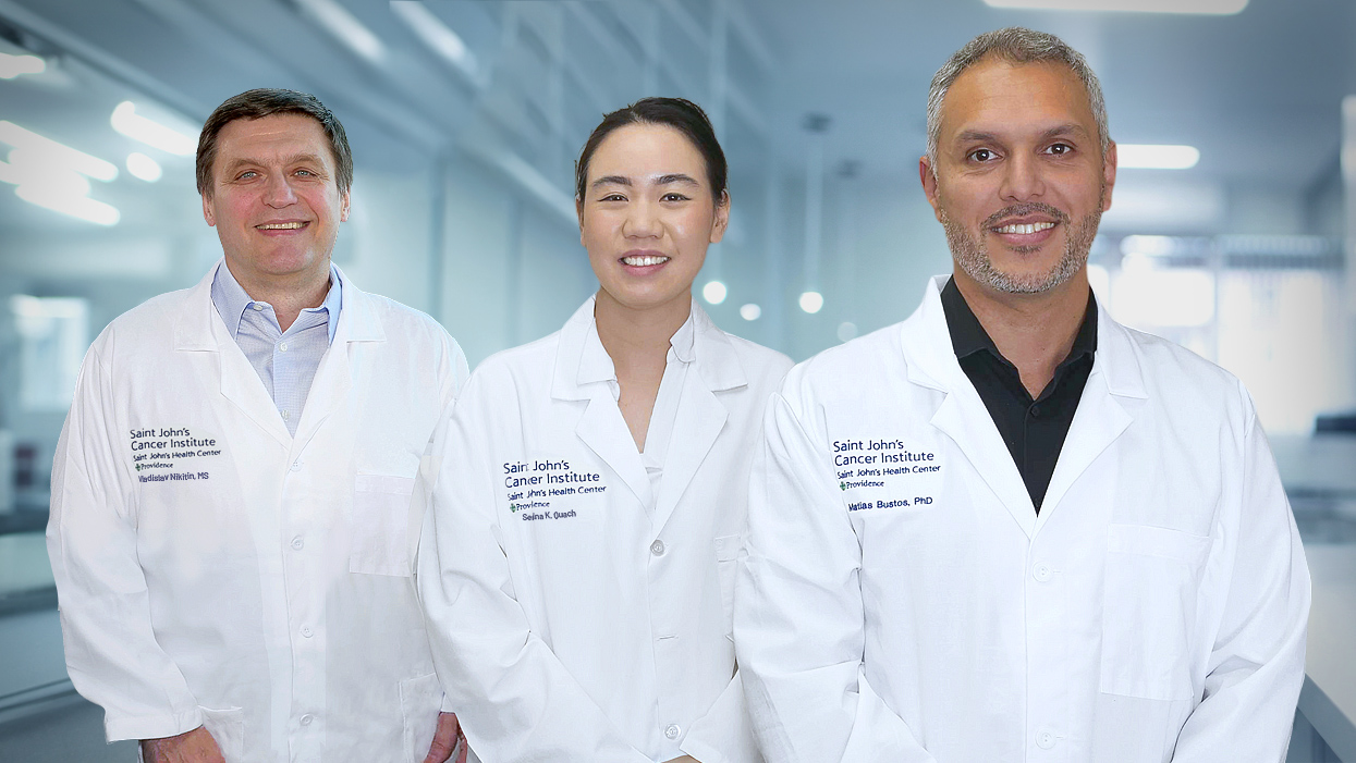
The Translational and Precision Medicine (TPM) Lab is dedicated to accelerating scientific discoveries into meaningful clinical applications that improve the diagnosis, risk assessment, and management of cancer patients.
Leveraging the extensive Saint John’s Cancer Institute Biorepository—including well‑annotated human tissues and biofluids (plasma, serum, and urine)—our team integrates next‑generation sequencing, spatial tumor profiling, and advanced computational tools to uncover biomarkers and molecular signatures associated with clinical application.
Founded in October 2025, the TPM Lab focuses on identifying translational research opportunities that directly enhance patient care, promote precision oncology, and support early intervention strategies across solid tumors and metastatic disease.
Current Projects
Developing Non‑Invasive Biomarkers for Early Detection and Monitoring of Renal Cell Carcinoma
Project Leads
PIs: Dr. Jennifer Linehan and Dr. Matias Bustos
Research Overview
The current study focuses on the development and validation of circulating microRNA (cfmiR) biomarkers capable of detecting and monitoring renal cell carcinoma (RCC) using non‑invasive liquid biopsy approaches. The project integrates expertise from the Department of Translational and Precision Medicine and the Urologic Oncology Department at Saint John’s Cancer Institute (SJCI), and the collaboration with Kidney Cancer Program at UCLA under Dr. Brian Shuch.
Clinical Need and Rationale
Kidney cancer remains a major clinical challenge: approximately 90,000 individuals are diagnosed annually in the U.S., and an estimated 20% will die from the disease. Currently, no blood‑ or urine‑based biomarkers exist for RCC. Imaging modalities such as MRI, ultrasound, and CT frequently detect renal masses incidentally, but cannot reliably differentiate malignant from benign lesions, nor predict tumor aggressiveness or recurrence risk. Thus, the development of sensitive, specific, and non‑invasive biomarkers could dramatically improve early detection, treatment stratification, and long-term surveillance for RCC patients.
Prior Findings
In a previous investigation (Bustos et al., Clinical Chemistry, 2024), the research team performed a comprehensive comparison of cfmiR profiles obtained from plasma and urine with matched RCC tumor tissues. Using a Next‑Generation Sequencing (NGS) platform assessing 2,083 microRNAs per sample, the study evaluated 221 biospecimens collected from 55 RCC patients. Key findings included:
- Identification of cfmiR signatures in plasma and urine that were consistently recapitulated in corresponding tumor tissues.
- Validation of these signatures against microRNA datasets from The Cancer Genome Atlas (TCGA) and additional published RCC-associated miRs.
- Demonstration that a specific panel of cfmiRs shows potential as a non‑invasive diagnostic biomarker set for RCC.
These pilot data provide the foundation for expanded clinical validation.
Current Study Aims
Supported by the PHASE ONE Foundation (2025–2027), the current study seeks to rigorously evaluate the clinical utility of cfmiRs as diagnostic and prognostic tools for RCC. The three primary aims are:
- Diagnostic Validation: Expand the RCC cohort to define a reproducible cfmiR signature capable of accurately distinguishing RCC from healthy controls and from benign renal conditions. Subtype discrimination (e.g., clear cell, papillary, chromophobe RCC) will also be explored.
- Prognostic Assessment: Determine whether specific cfmiRs correlate with disease aggressiveness, including pathological stage, tumor biology, and risk of recurrence.
- Surveillance Utility: Examine whether cfmiR profiles can serve as reliable markers for postoperative monitoring, enabling earlier detection of recurrence compared to imaging alone.
Methodological Approach
The study incorporates a pre-enrichment workflow combined with NGS-based miR profiling across matched blood, urine, and tumor tissue samples. A cohort of 65 RCC patients will be enrolled, along with healthy donors and individuals with benign kidney diseases to provide robust comparison groups.
Reference
Bustos MA, Gottlieb J, Choe J, Suyeon R, Lin SY, Allen WM, Krasne DL, Wilson TG, Hoon DSB, Linehan JA. Diagnostic miRNA Signatures in Paired Tumor, Plasma, and Urine Specimens From Renal Cell Carcinoma Patients. Clin Chem. 2024 Jan 4;70(1):261–272. doi: 10.1093/clinchem/hvad133. PMID: 37791385.
TPM Offering
The TPM lab offers services for digital pathology analysis, including slide scanning and analysis. The current process involved three main steps:
- Acquire a new tissue section that has been stained (H&E or any specific markers using IHC)
- Scan the slides
- Perform image analysis using different algorithms and AI-driven tools based on imaging recognition.
The TPM lab also provides specialized services in molecular profiling and bioinformatic interpretation, supporting clinical research programs at Saint John’s Cancer Institute and partner institutions.
See Services below.
Services
Digital Pathology Analysis
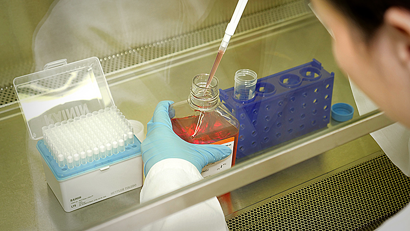
Digital pathology enables scalable, quantitative, and reproducible analysis of tumor tissue using a single slide stained with hematoxylin and eosin (which is normally used as the standard of care in the Pathology Dept). When combined with next-generation sequencing-based analysis (genomic, transcriptomic, epigenomic) and spatial profiling, it allows researchers to:
- Identify subtle histological patterns undetectable by visual inspection
- Correlate tissue features with genomic variants, gene expression, or immune profiles
- Stratify patients based on the features identified to predict response to therapy, monitor post‑treatment tumor evolution, and mechanisms of resistance
This integrative approach accelerates biomarker discovery and supports the development of personalized therapeutic strategies. Our digital pathology pipeline supports high‑quality imaging, quantitative tissue analysis, and AI‑driven biomarker discovery.
The workflow includes:
Tissue Preparation & Staining
- Standard H&E staining
- Immunohistochemistry (IHC) for tumor, immune, and stromal markers
- Multiplex IHC / IF (optional future expansion
Whole‑Slide Imaging (WSI)
- High‑resolution scanning of glass slides with brightfield or fluorescence capability
- Generation of standardized, compatible digital formats (SVS, NDPI, TIFF)
- Quality control for image clarity, sharpness, and artifact detection
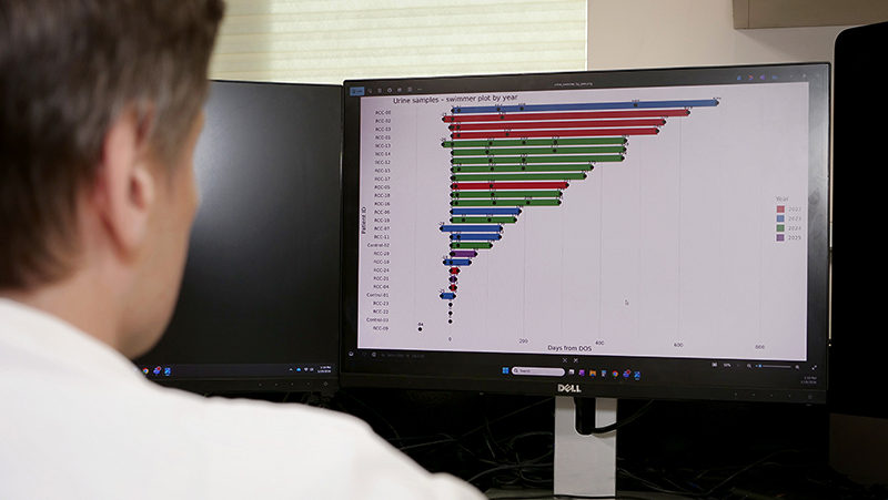
AI‑Powered Image Analysis Using machine‑learning and deep‑learning algorithms, we provide:
- Automated cell segmentation and phenotyping
- Quantification of immune infiltration, stromal content, necrosis, and tumor architecture
- Detection of spatial relationships between tumor and immune cells
- Identification of predictive markers associated with treatment resistance or metastasis. The TPM Lab incorporates state‑of‑the‑art digital pathology tools and algorithmic analysis pipelines to support translational research studies.
Data Integration & Reporting
- Comprehensive analytical reports with visual overlays, heatmaps, and region‑specific metrics
- Integrated interpretation with molecular data (NGS, spatial profiling, miRNA/mRNA expression)
- Customized data outputs for IRB‑approved clinical or translational research projects