


How to Spot Skin Cancer: Recognizing Changes in Your Skin
Skin cancer can appear as a new mole, a changing spot, a persistent sore, or a growth that looks different from the surrounding skin. Some skin cancers are darkly pigmented, while others are pink, red, skin-colored, or nearly colorless. Knowing what to look for can help you recognize suspicious changes and seek medical evaluation before a potential problem becomes more advanced.
Skin cancer is the most commonly diagnosed type of cancer in the United States. Basal cell carcinoma and squamous cell carcinoma are the most common forms, while melanoma and Merkel cell carcinoma are less common but can be more aggressive. Although many skin cancers develop in areas exposed to ultraviolet (UV) radiation, they can also appear in places that receive little sunlight.
Most skin spots and growths are not cancerous. However, appearance alone cannot reliably determine whether a lesion is harmless or malignant. A dermatologist or other qualified healthcare professional can examine concerning changes and determine whether additional evaluation or a biopsy is appropriate.
What Does Skin Cancer Look Like?
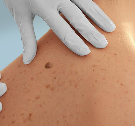
Skin cancer does not have a single appearance. Its color, shape, texture, and growth pattern depend on the type of cancer, its location, and individual skin characteristics. Some lesions change noticeably over weeks or months, while others develop more gradually.
Common warning signs include a new or changing mole, a sore that does not heal, a spot that repeatedly bleeds or crusts, a rough or scaly patch, or a growth that looks noticeably different from other spots on your skin.
Basal Cell Carcinoma (BCC)
Basal cell carcinoma is the most common skin cancer. It frequently develops on areas exposed to the sun, including the face, ears, scalp, neck, and arms, although it can occur elsewhere. BCC may appear as a shiny or pearly bump, a pink or skin-colored growth, a flat patch, or a sore that repeatedly heals and reopens. Some lesions contain visible small blood vessels or areas of brown or darker pigmentation.
Basal cell carcinomas generally grow slowly and rarely spread to distant parts of the body. However, untreated tumors can invade surrounding tissue and cause significant local damage.
Learn more about Basal Cell Carcinoma: Symptoms, Diagnosis, and Treatment.
Squamous Cell Carcinoma (SCC)
Squamous cell carcinoma often develops in areas with cumulative sun exposure, including the face, ears, lips, neck, and backs of the hands. It may appear as a rough or scaly patch, a firm raised growth, a thickened or wart-like area, or a sore that crusts, bleeds, or does not heal. Its appearance can vary across skin tones, including red, brown, darkly pigmented, or skin-colored lesions.
Some squamous cell carcinomas can grow deeply or spread to nearby lymph nodes or other tissues. Early evaluation is important, particularly for rapidly growing or persistent lesions.
Learn more about Squamous Cell Carcinoma of the Skin.
Melanoma
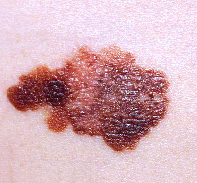
Melanoma develops from melanocytes, the cells that produce skin pigment. It may arise in an existing mole or appear as an entirely new lesion. Melanoma often presents as a brown or black spot with uneven color or irregular borders, but some melanomas are pink, red, or relatively unpigmented.
Melanoma is especially concerning because it can invade deeper tissue and spread to other parts of the body. Recognizing changes early can help patients obtain a timely diagnosis and appropriate treatment.
Learn more about Melanoma Skin Cancer: Symptoms, Stages, and Treatment.
Merkel Cell Carcinoma (MCC)
Merkel cell carcinoma is a rare, aggressive skin cancer that often appears as a firm, rapidly growing bump or nodule. The growth may be red, pink, violet, or skin-colored and is often painless. These tumors commonly develop on sun-exposed skin but can occur in other locations.
Because Merkel cell carcinoma can grow and spread quickly, a new or rapidly enlarging unexplained nodule should be evaluated promptly.
Learn more about Merkel Cell Carcinoma: Signs, Diagnosis, and Treatment.
How to Recognize Melanoma: The ABCDE Warning Signs
The ABCDE method is a useful way to identify features that may be associated with melanoma. It helps people notice suspicious changes in moles and pigmented spots, but it is not a diagnostic test. Some melanomas do not display all five warning signs, and some harmless moles may have unusual features.
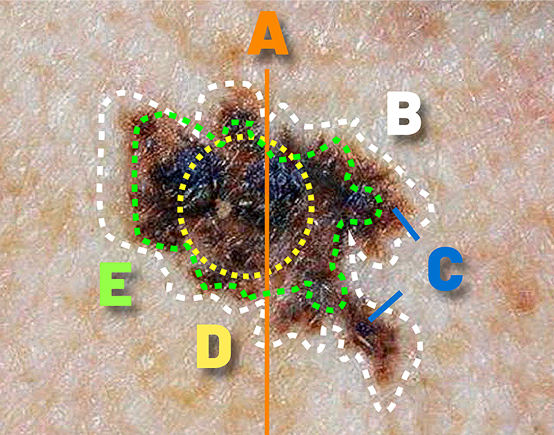
- A — Asymmetry: One half of a mole or spot looks different from the other half.
- B — Border: The edges are uneven, notched, blurred, or irregular.
- C — Color: The lesion has uneven pigmentation or multiple colors, including brown, black, red, white, or blue.
- D — Diameter: Melanomas are often larger than approximately 6 millimeters when diagnosed, but they can also be smaller.
- E — Evolving: The spot changes in size, shape, color, elevation, or symptoms, including new itching or bleeding.
Among these features, evolving or changing is particularly important. A spot that is becoming noticeably different should be evaluated even if it is smaller than a pencil eraser or does not appear especially dark.
The Ugly Duckling Sign
Most people have moles that share a similar appearance. The ugly duckling sign refers to a spot that looks noticeably different from the person’s other moles in color, size, shape, or overall pattern.
A distinctive lesion is not necessarily cancerous, but it deserves closer attention, particularly if it is new or changing. Comparing spots with other marks on your skin can sometimes be more useful than examining a single mole in isolation.
Can Melanoma Be Pink, Red, or Skin-Colored?
Yes. Although melanoma is often associated with darkly pigmented moles, some melanomas contain little visible pigment. These are sometimes called amelanotic melanomas and may appear pink, red, or similar to the surrounding skin.
For this reason, a new or changing growth should not be dismissed simply because it does not resemble a typical dark mole.
Can Skin Cancer Be Itchy, Painful, or Bleeding?
Skin cancer may cause itching, tenderness, bleeding, crusting, or pain, but some lesions have no noticeable symptoms. A persistent sore that does not heal, a growth that repeatedly scabs and reopens, or a spot that bleeds without an obvious injury should be evaluated by a healthcare professional.
Many noncancerous skin conditions can also cause these symptoms. Eczema, irritation, infections, and other dermatologic conditions may produce itching or scaling. However, recurring symptoms in the same location or a lesion that continues to change should not be ignored.
It is also important to recognize that skin cancer can develop without discomfort. Waiting for pain, itching, or bleeding may delay the assessment of an otherwise suspicious lesion.
Age Spots, Sunspots, and Skin Cancer: How Can You Tell the Difference?
Age spots, also known as solar lentigines or sunspots, are generally harmless areas of increased pigmentation associated with cumulative sun exposure. They commonly appear on the face, hands, shoulders, and other areas frequently exposed to sunlight, particularly as people get older.
Typical age spots are flat, have relatively uniform pigmentation, and often remain stable over time. However, their appearance can overlap with certain melanomas and other pigmented skin conditions. Seborrheic keratoses, which are common noncancerous growths, can also resemble skin cancer.
A spot that changes in color, shape, size, or texture, develops an irregular border, or looks different from surrounding spots should be professionally evaluated. Do not assume that a new dark mark is simply an age spot, especially if its appearance is evolving.
Age spots do not generally require medical treatment, but cosmetic lightening or removal should be considered only after the lesion has been properly assessed. Attempting to fade an undiagnosed spot could delay the recognition of skin cancer.
Where Can Skin Cancer Develop on the Body?
Many skin cancers occur on sun-exposed areas, including the face, ears, scalp, neck, shoulders, arms, and hands. However, melanoma and other skin cancers may also arise on the back, legs, feet, and parts of the body that are usually covered by clothing.
Skin cancer can affect people of every skin tone. Although some types are more common in people with lighter skin, darker skin does not eliminate the possibility of skin cancer. Some cancers in people with darker skin may occur in less sun-exposed locations and can be overlooked.
Don’t Overlook Your Palms, Soles, and Nails
Certain melanomas, including acral lentiginous melanoma, may develop on the palms of the hands, soles of the feet, or beneath the nails. Warning signs can include a new or changing dark spot, irregular pigmentation, a persistent lesion, or a dark band beneath a fingernail or toenail that changes over time.
Not every dark nail streak is melanoma. However, unexplained nail pigmentation, particularly if it is new, changing, irregular, or extends onto surrounding skin, should be evaluated rather than assumed to be a bruise or harmless discoloration.
Skin Changes That May Be Easier to Miss
When checking your skin, remember areas such as the scalp, behind the ears, between the toes, the lower back, and the backs of the legs. Some spots are difficult to see without a mirror or assistance from another person.
Any persistent or unusual lesion in an area that receives little sunlight deserves the same attention as a suspicious spot on the face or arms.
How to Check Your Skin for Suspicious Changes
Becoming familiar with your skin can help you recognize new spots or changes in existing moles. A skin self-check does not replace professional evaluation, but it can help you identify concerns to discuss with a dermatologist or another qualified clinician.
Use a well-lit room, a full-length mirror, and a hand mirror when needed. Look for changing moles, unusual spots, persistent sores, and areas that appear different from the surrounding skin.
- Examine your face, ears, and neck. Include your lips and areas around the hairline.
- Check your scalp. Part your hair to examine the skin beneath it, or ask someone to help.
- Look at your hands and arms. Check your palms, fingernails, forearms, upper arms, and underarms.
- Examine your chest, abdomen, and back. Use mirrors to inspect areas you cannot see directly.
- Check your legs and feet. Include the backs of the legs, soles, between the toes, and toenails.
- Notice changes over time. If a spot is new, evolving, bleeding, or otherwise concerning, arrange a professional examination.
For individuals with numerous moles or lesions requiring surveillance, a clinician may recommend photography or other methods to document and compare changes. The frequency of professional examinations should be individualized according to medical history, risk factors, and previous skin findings.
Learn more about the Pigmented Skin Lesion Clinic and Skin Cancer Evaluation at Saint John’s.
Who Is at Increased Risk for Skin Cancer?

Skin cancer can develop in anyone, but certain characteristics and exposures may increase risk. These include a personal history of skin cancer, a family history of melanoma, numerous or atypical moles, significant lifetime ultraviolet exposure, blistering sunburns, indoor tanning, and a weakened immune system.
People with lighter skin, light-colored eyes, or hair that burns easily may have greater susceptibility to UV-related skin damage. However, individuals with darker skin tones can also develop melanoma and other skin cancers, including lesions in areas that receive minimal sunlight.
Some inherited conditions are associated with increased melanoma risk. Patients with a strong family history or multiple affected relatives may benefit from a discussion about individualized surveillance and, in selected cases, genetic counseling.
Risk factors help guide evaluation, but they do not determine whether an individual spot is cancerous. A concerning lesion should be assessed even when the person has no known risk factors.
When Should You See a Doctor About a Skin Spot?
Arrange an evaluation if you notice a mole that is changing, a new spot that looks different from your other moles, a sore that does not heal, unexplained bleeding, or a growth that is rapidly enlarging. A suspicious lesion should not be monitored indefinitely at home while waiting for additional symptoms to develop.
A healthcare professional may examine the lesion with magnification or dermoscopy and review your medical history and risk factors. If cancer is suspected, a skin biopsy may be recommended to establish the diagnosis.
For people without symptoms or a history suggesting elevated risk, recommendations about routine clinician-performed skin screening are less definitive. The U.S. Preventive Services Task Force has found insufficient evidence to recommend for or against routine visual skin screening in the general asymptomatic population. This does not mean that concerning lesions should go unevaluated.
People with previous skin cancer, suspicious lesions, or other important risk factors may need individualized follow-up. Your dermatologist or treating clinician can recommend an appropriate examination and surveillance schedule.
What Happens During a Skin Lesion Evaluation?
An examination typically includes a review of the lesion’s history, changes you have noticed, relevant risk factors, and inspection of the affected skin. Depending on the findings, a clinician may use dermoscopy, compare previous photographs, recommend monitoring, or arrange a biopsy.
A biopsy allows a pathologist to examine tissue under a microscope and can establish whether the lesion is cancerous. Not every suspicious spot requires a biopsy, and not every biopsy reveals cancer.
Specialized Evaluation at the Pigmented Skin Lesion Clinic
The Pigmented Skin Lesion Clinic at Providence Saint John’s Health Center provides evaluation of suspicious moles, pigmented lesions, and other concerning skin changes. The program includes clinical assessment, dermoscopy, photographic monitoring when appropriate, and coordination with pathology and melanoma specialists when additional care is needed.
Patients with complex or confirmed skin cancers can be referred for further evaluation through the Borstein Family Foundation Melanoma Program and other clinical services at Saint John’s Cancer Institute.
Learn more about Skin Cancer Screening and the Pigmented Skin Lesion Clinic.
How Can You Reduce Your Risk of Skin Cancer?
Although no prevention strategy eliminates every skin cancer risk, reducing unnecessary ultraviolet exposure is an important way to protect your skin. Sun-protective habits are valuable throughout life, including during childhood and adulthood.
- Use sunscreen: Choose broad-spectrum, water-resistant sunscreen with SPF 30 or higher, applying it to exposed skin and reapplying approximately every two hours outdoors and after swimming or sweating.
- Seek shade: Limit prolonged direct sun exposure, particularly around midday when UV radiation is often strongest.
- Wear protective clothing: Consider wide-brimmed hats, UV-protective sunglasses, and clothing with adequate sun protection.
- Avoid indoor tanning: Tanning beds expose skin to ultraviolet radiation and increase the risk of skin cancer.
- Pay attention to the UV Index: Check local UV conditions and adjust your protective measures accordingly.
Sun protection remains important on cloudy days because ultraviolet radiation can still reach the skin. People who spend extended periods outdoors may need additional protective measures based on the duration and intensity of exposure.
For more detailed guidance, read Precautions to Take as the UV Index Rises.
Recognizing Skin Cancer Early and Taking the Next Step
Learning how to spot skin cancer begins with recognizing what is normal for your skin and paying attention to changes. A new or evolving mole, persistent sore, unusual growth, or lesion that looks different from surrounding spots may need professional evaluation. Because skin cancers vary in appearance and can occur anywhere on the body, a qualified clinician—not a visual checklist alone—is essential for determining whether a suspicious lesion requires further testing.
The Pigmented Skin Lesion Clinic at Providence Saint John’s Health Center provides evaluation of concerning moles and skin changes, with access to specialized melanoma care when appropriate. To learn more about skin cancer evaluations or schedule an appointment, call 310-829-8317.
Sources & Further Reading
- American Academy of Dermatology — ABCDE Warning Signs of Melanoma
- American Academy of Dermatology — How to Perform a Skin Self-Exam
- American Academy of Dermatology — Finding Skin Cancer in Darker Skin Tones
- American Academy of Dermatology — Age Spots and the Importance of Evaluation
- U.S. Preventive Services Task Force — Skin Cancer Screening Recommendation (2023)
- American Cancer Society — Squamous Cell Skin Cancer Signs and Symptoms
- Saint John’s Cancer Institute — Pigmented Skin Lesion Clinic
- Saint John’s Cancer Institute — Skin Cancer Conditions