We are committed to diagnosing and treating liver cancer, always with a goal of developing more effective therapies.

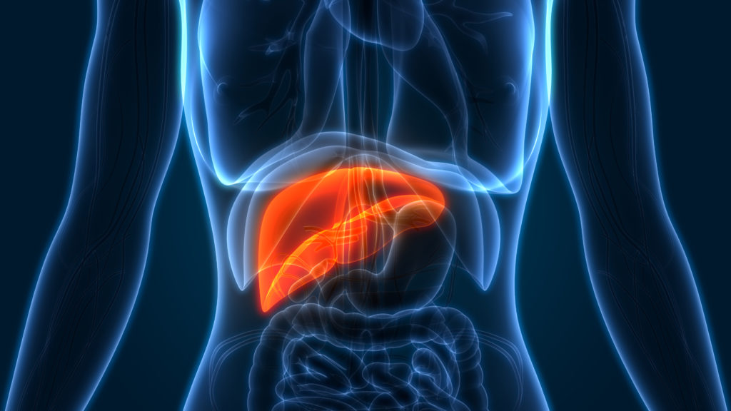
We evaluate many patients each year with primary liver cancer, and use every means at our disposal to treat hepatocellular carcinoma, which arises in liver cells known as hepatocytes, and cholangiocarcinoma that begins in the bile ducts within the liver.

We also treat many types of benign liver tumors well as a rare type of liver cancer called fibrolamellar-hepatocellular carcinoma.

In order to reach a diagnosis for liver, biliary, or pancreatic disorders, we take a thorough medical history in order to be as accurate as possible. We’ll ask questions and make note of any symptoms you may have experienced as well any other pertinent information.

A physical examination is also done to help assess the problem more completely.

3D Image of a Liver

Diagnosing liver cancer or metastatic liver cancer may include one or more of the following tests/procedures:

Abdominal ultrasound or sonography:

This diagnostic imaging technique uses high-frequency sound waves to create an image of your internal organs such as the liver, spleen, and kidneys and to assess blood flow through various vessels.

Computed tomography scan (CT or CAT scan):

This diagnostic imaging procedure uses a combination of X-rays and computer technology to produce very detailed images – often called slices – of the body, including the organs.

Endoscopic retrograde cholangiopancreatography (ERCP):

This procedure allows the gastroenterologists at Providence Saint Johns to diagnose and treat problems in the liver, gallbladder, bile ducts, and pancreas. The procedure combines X-ray and the use of an endoscope, a long, flexible, lighted tube. The scope is guided through the patient’s mouth and throat, then through the esophagus, stomach, and duodenum. The doctor can examine the inside of these organs and detect any abnormalities. A tube is then passed through the scope, and a dye is injected which will allow the internal organs to appear on an X-ray. Stents or shunts can be placed to relieve blocked bile ducts.

Esophagogastroduodenoscopy (also called EGD or upper endoscopy):

This allows your doctor to examine the inside of the esophagus, stomach, and duodenum. A thin, flexible, lighted tube, called an endoscope, is guided into the mouth and throat, then into the esophagus, stomach, and duodenum. The endoscope allows the doctor to view the inside of this area of the body, as well as to insert instruments through a scope for the removal of a sample of tissue for biopsy if a biopsy is deemed necessary.

Laparoscopy:

This diagnostic test uses a viewing tube with a lens or camera and a light on the end, which is inserted through a small incision in the abdomen, to examine the contents of the abdomen and remove tissue samples.

Liver biopsy:

This procedure takes tissue samples from the liver with a needle or during surgery for examination under a microscope.

Liver function tests:

This is a series of special blood tests that can determine if the liver is functioning properly.

Magnetic resonance cholangiopancreatography (MRCP):

This is a special type of MRI that uses radio waves and magnets to obtain pictures of the bile ducts and internal organs.

Magnetic Resonance Imaging (MRI):

This imaging modality uses a powerful to find tumors in the liver. This is often more sensitive than CT scans for primary liver cancer especially in patients with cirrhosis.

Percutaneous transhepatic cholangiography (PTC):

A needle is introduced through the skin and into the liver where a dye is deposited so the bile duct structures can then be viewed by X-ray.

X-ray:

This diagnostic test uses invisible electromagnetic energy beams to produce images of internal tissues, bones, and organs onto film.