What Is Stomach Cancer?
Stomach cancer, also called gastric cancer, develops when abnormal cells grow uncontrollably within the stomach. Most stomach cancers begin in the glandular cells lining the stomach and are known as adenocarcinomas. Early-stage stomach cancer may cause few or no noticeable symptoms, while more advanced disease can interfere with digestion, cause bleeding, or spread beyond the stomach. Treatment depends on the type, location, and stage of the cancer, as well as the patient’s overall health and individual needs.
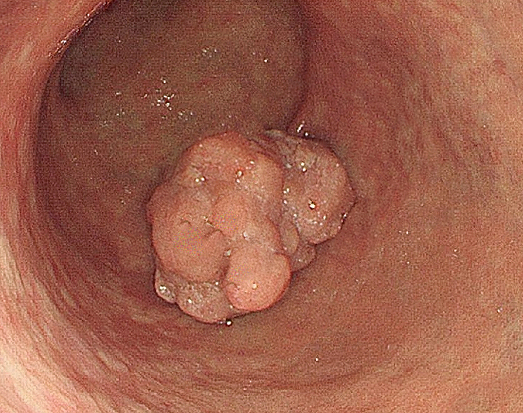
The stomach is a muscular organ located between the esophagus and small intestine. It stores food, produces acid and digestive substances, and helps begin the process of digestion. Its inner lining contains glandular cells that can undergo changes over time, sometimes progressing from chronic inflammation to precancerous changes and eventually cancer. However, not every stomach ulcer or polyp is precancerous, and many stomach conditions are unrelated to cancer.
At Saint John’s Cancer Institute in Santa Monica, multidisciplinary evaluation brings together specialists in surgical oncology, medical oncology, radiation oncology, gastroenterology, diagnostic imaging, pathology, and supportive care. This coordinated approach helps determine the extent of disease, identify appropriate treatment options, and support patients and families throughout their care.
Causes and Types of Stomach Cancer
Stomach cancer develops through changes that allow cells to grow and divide abnormally. An important risk factor for many stomach cancers is chronic infection with Helicobacter pylori (H. pylori), a bacterium that can cause persistent inflammation of the stomach lining. Other factors include tobacco use, certain dietary patterns, autoimmune gastritis, and inherited cancer-predisposition syndromes. Some cancers near the junction of the stomach and esophagus are associated with obesity and gastroesophageal reflux disease.
Most stomach cancers are adenocarcinomas, but they can differ according to where they develop and how their cells grow. These distinctions are important because cancers in different locations may require different diagnostic, surgical, and medical treatment approaches.
Non-Cardia Stomach Cancer
Non-cardia gastric cancer develops in areas of the stomach away from its junction with the esophagus. Many cases are associated with long-standing H. pylori infection, which can damage the stomach lining through chronic inflammation. In some patients, this process leads to atrophic gastritis, intestinal metaplasia, dysplasia, and eventually cancer. H. pylori infection is common worldwide, but most infected individuals never develop stomach cancer. Testing and treating the infection can reduce cancer risk, particularly when performed before extensive precancerous changes have developed.
Diffuse-Type Stomach Cancer
Diffuse-type gastric adenocarcinoma is characterized by cancer cells that infiltrate the stomach wall rather than forming a clearly defined mass. Some tumors contain signet-ring cells, and extensive involvement may cause the stomach wall to become thickened and rigid, a condition known as linitis plastica. Because these cancers may spread beneath an apparently normal-looking stomach lining, diagnosis can be challenging and sometimes requires additional or deeper tissue sampling. A small proportion are associated with inherited mutations, particularly involving the CDH1 gene.
Cardia and Gastroesophageal Junction Cancer
The cardia is the upper portion of the stomach near the gastroesophageal junction (GEJ), where the esophagus meets the stomach. Cancers in this region may involve stomach tissue, the lower esophagus, or both. Their classification and treatment planning depend on the location of the tumor’s center and how far it extends across the junction. Risk factors for some proximal gastric and GEJ adenocarcinomas include obesity and chronic reflux, although these relationships differ from those of H. pylori-associated non-cardia cancers.
For additional information about cancers involving the lower esophagus and gastroesophageal junction, visit our Esophageal Cancer page.
Less Common Stomach Tumors

Although adenocarcinoma accounts for most stomach cancers, several other types of tumors can arise in or involve the stomach. These conditions differ in their biology, likelihood of spread, and treatment requirements.
Gastric Lymphoma
Gastric lymphoma develops from immune system cells within the stomach. Types include mucosa-associated lymphoid tissue (MALT) lymphoma and more aggressive lymphomas, such as diffuse large B-cell lymphoma. Some MALT lymphomas are associated with H. pylori infection and may respond to eradication of the bacteria. Other lymphomas require treatments such as immunotherapy, chemotherapy, or radiation rather than stomach surgery.
Gastrointestinal Stromal Tumor (GIST)
Gastrointestinal stromal tumors arise from specialized cells associated with the muscular wall of the digestive tract. They are biologically distinct from stomach adenocarcinomas and may be classified according to tumor size, location, growth rate, and molecular characteristics. Treatment can include surgical removal and selected targeted medications, depending on the tumor’s behavior and genetic alterations.
Gastric Neuroendocrine Tumors
Gastric neuroendocrine tumors develop from hormone-producing cells in the stomach lining. Some are associated with chronic atrophic gastritis or specific hormone-related conditions, while others occur sporadically. Their behavior ranges from relatively indolent to aggressive, making accurate classification, grading, and staging important when determining whether surveillance, endoscopic removal, surgery, or systemic treatment is appropriate.
Signs and Symptoms of Stomach Cancer
Early stomach cancer may not produce recognizable symptoms. When symptoms develop, they can resemble those of common digestive conditions, including gastritis, peptic ulcers, and acid reflux. Persistent symptoms deserve medical evaluation, particularly when accompanied by unexplained weight loss, anemia, or evidence of gastrointestinal bleeding.

Possible signs and symptoms of stomach cancer include:
- Persistent upper abdominal discomfort or pain
- Feeling full after eating only a small amount of food
- Unexplained weight loss or loss of appetite
- Nausea, vomiting, or ongoing indigestion
- Difficulty swallowing, particularly when the tumor is near the gastroesophageal junction
- Fatigue, weakness, or anemia related to chronic blood loss
- Black, tarry stools or vomiting blood
- Abdominal swelling or other symptoms associated with more advanced disease
These symptoms do not necessarily mean a person has stomach cancer. However, progressive difficulty swallowing, unexplained weight loss, persistent vomiting, or symptoms of bleeding should not be ignored. Vomiting blood, substantial gastrointestinal bleeding, or signs of severe blood loss require urgent medical attention.
How Is Stomach Cancer Diagnosed?
Diagnosing stomach cancer begins with a medical history, review of symptoms, and physical examination. If a patient has persistent upper abdominal discomfort, difficulty swallowing, unexplained weight loss, anemia, or gastrointestinal bleeding, additional testing may be recommended. Upper endoscopy with biopsy is the principal method for confirming stomach cancer. Imaging and other procedures help determine its extent and guide treatment planning.
Upper Endoscopy (EGD) and Biopsy
Esophagogastroduodenoscopy (EGD), also called upper endoscopy, uses a thin, flexible tube with a camera to examine the lining of the esophagus, stomach, and first part of the small intestine. During the procedure, a gastroenterologist can identify suspicious areas and obtain tissue samples for examination by a pathologist. A biopsy is necessary to confirm most stomach cancer diagnoses and determine the type of tumor. Tissue may also be evaluated for H. pylori infection and molecular biomarkers that could influence treatment decisions.
Endoscopic Ultrasound (EUS)

Endoscopic ultrasound combines an endoscope with a small ultrasound probe to create detailed images of the stomach wall and surrounding structures. It is particularly useful in selected patients with potentially localized disease because it can help determine how deeply a tumor has invaded and whether nearby lymph nodes appear abnormal. EUS may also guide needle sampling when additional tissue is required.
Computed Tomography (CT)
CT scanning produces detailed cross-sectional images of the body and is commonly used after a stomach cancer diagnosis to evaluate disease extent. Imaging of the chest, abdomen, and pelvis may identify enlarged lymph nodes, involvement of nearby structures, or suspected spread to organs such as the liver. CT findings help determine whether surgery may be appropriate and whether additional staging procedures are needed.
Staging Laparoscopy
For selected patients with potentially resectable but more extensive stomach cancer, staging laparoscopy may be performed before definitive surgery. Through small abdominal incisions, a surgeon uses a camera to examine the abdominal cavity for cancer deposits that may not appear on conventional imaging. Fluid may also be collected through peritoneal washings to check for microscopic cancer cells. These findings can significantly affect treatment recommendations.
Additional Imaging and Laboratory Tests
Other tests may be used selectively. PET/CT can help investigate suspected cancer spread in certain patients, although some stomach cancers, particularly diffuse-type tumors, may not show strong PET activity. An upper GI series using swallowed barium can reveal structural abnormalities but is not generally preferred over endoscopy for diagnosing stomach cancer. Blood tests can identify anemia and evaluate organ function, while stool testing may detect hidden gastrointestinal bleeding. These tests support evaluation but do not independently establish a stomach cancer diagnosis.
Stomach Cancer Stages
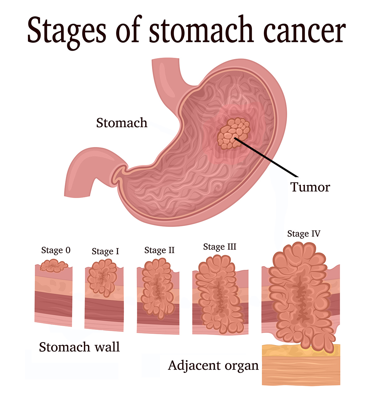
Staging describes how far stomach cancer has developed and whether it has spread beyond its original location. For gastric adenocarcinoma, the American Joint Committee on Cancer (AJCC) TNM system considers the depth of tumor invasion, the number of regional lymph nodes involved, and whether distant metastases are present. Clinical staging uses findings from imaging, endoscopy, biopsy, and other examinations. When surgery is performed, examination of the removed tissue may provide additional staging information.
Understanding the TNM System
T — Primary Tumor: Describes how deeply the cancer has grown into the layers of the stomach wall or whether it extends into nearby structures.
N — Regional Lymph Nodes: Describes the extent of cancer involvement in nearby lymph nodes. The number of involved lymph nodes helps determine the stage.
M — Distant Metastasis: Indicates whether cancer has spread to distant sites, including the liver, peritoneum, or distant lymph nodes.
Stage 0 — Carcinoma in Situ
Stage 0 refers to abnormal cells confined to the innermost lining of the stomach without invasion into deeper tissues. These changes may be identified during endoscopy and, in selected cases, treated with endoscopic removal.
Stage I — Early Invasive Cancer
Stage I generally describes cancer that has invaded the stomach wall but remains relatively limited in extent, with no or limited nearby lymph node involvement, depending on tumor depth. Some very early cancers may be suitable for endoscopic treatment, while others require surgery and lymph node evaluation.
Stage II — More Extensive Local Disease
Stage II includes certain combinations of deeper invasion into the stomach wall and regional lymph node involvement. Although the cancer has not spread to distant organs, treatment often involves surgery combined with systemic therapy, depending on the patient’s clinical stage and other factors.
Stage III — Locally Advanced Cancer
Stage III generally indicates more extensive involvement of the stomach wall, nearby structures, or regional lymph nodes, without distant metastasis. Treatment may include chemotherapy before and after surgery, with other approaches considered according to tumor characteristics and whether complete surgical removal is feasible.
Stage IV — Metastatic Stomach Cancer
Stage IV means stomach cancer has spread to distant organs or tissues. Common sites include the liver, peritoneum, and distant lymph nodes. Treatment generally focuses on systemic therapies selected according to cancer characteristics, molecular biomarkers, and patient needs, together with supportive care. Local procedures may also help manage bleeding, obstruction, or other symptoms.
These stage descriptions are general summaries rather than a substitute for the complete TNM stage-grouping criteria. The stage may be refined after surgery or reassessment following treatment. Staging for gastric lymphoma, GIST, and gastric neuroendocrine tumors follows different classification systems.
Risk Factors for Stomach Cancer
Stomach cancer develops through a combination of environmental, infectious, genetic, and other factors. Having a risk factor does not mean a person will develop cancer, and some patients are diagnosed without any clearly identifiable risk factors. Understanding the major associations can help individuals discuss prevention, appropriate evaluation, and family risk with their healthcare team.
Helicobacter pylori Infection
Chronic infection with Helicobacter pylori (H. pylori) is a major established risk factor for non-cardia gastric adenocarcinoma. The bacterium can produce long-standing inflammation of the stomach lining, which in some individuals leads to atrophic gastritis and precancerous changes. Most infected people do not develop stomach cancer, but identifying and treating the infection can reduce risk. Treatment typically involves a combination of antibiotics and acid-suppressing medication, followed by testing to confirm successful eradication.
Age, Sex, and Population Risk

Stomach cancer becomes more common with age and is diagnosed more frequently in men than women. In the United States, incidence also differs among population groups, reflecting a complex combination of H. pylori prevalence, geographic ancestry, environmental exposures, socioeconomic conditions, and access to preventive healthcare. These differences should be interpreted as population-level patterns rather than evidence that race itself causes stomach cancer.
Smoking, Diet, and Body Weight
Tobacco smoking increases stomach cancer risk and is a preventable contributor to several gastrointestinal cancers. Diets high in heavily salted or salt-preserved foods are also associated with increased risk, while diets that include a variety of fruits and vegetables may contribute to better overall health. Obesity is particularly associated with cancers near the upper stomach and gastroesophageal junction. Reducing tobacco exposure and maintaining healthy dietary and activity patterns can support cancer risk reduction, although no single food or lifestyle change eliminates the possibility of stomach cancer.
Chronic Gastritis and Other Stomach Conditions
Chronic atrophic gastritis and intestinal metaplasia are changes in the stomach lining associated with increased cancer risk in certain patients. Autoimmune gastritis, sometimes associated with pernicious anemia and vitamin B12 deficiency, is another recognized risk factor. Patients with these conditions may benefit from individualized discussion about H. pylori testing, endoscopic evaluation, and whether surveillance is appropriate.
Family History and Inherited Cancer Syndromes
Having a first-degree relative with stomach cancer can increase personal risk. A smaller proportion of gastric cancers are associated with inherited genetic syndromes, including hereditary diffuse gastric cancer involving CDH1 or CTNNA1, Lynch syndrome, and certain polyposis syndromes. CDH1-related hereditary diffuse gastric cancer is particularly important because affected families may also have an increased risk of lobular breast cancer.
Individuals with multiple relatives diagnosed with stomach cancer, stomach cancer at unusually young ages, or a family pattern suggestive of hereditary cancer may benefit from genetic counseling. Testing and surveillance recommendations depend on the specific family history and any identified inherited mutation.
Stomach Cancer Treatment Overview
Treatment for stomach cancer depends on the type and stage of the disease, its location within the stomach, the patient’s overall health, and the results of diagnostic and molecular testing. Some early cancers can be treated with endoscopic procedures or surgery, while more advanced cancers may require chemotherapy, radiation therapy, immunotherapy, targeted medications, or a combination of approaches. A multidisciplinary team helps determine the appropriate sequence of treatment and whether surgery offers a potential opportunity for cure.
Endoscopic Treatment for Early Stomach Cancer
Selected very early-stage stomach cancers that are confined to superficial layers and have a low risk of lymph node involvement may be removed during endoscopy. Techniques such as endoscopic submucosal dissection (ESD) allow specialists to remove certain lesions without removing part of the stomach. Careful evaluation of tumor size, depth, appearance, and pathology helps determine whether endoscopic treatment is appropriate or whether additional surgery is necessary.
Partial or Subtotal Gastrectomy
Partial gastrectomy removes the portion of the stomach containing the cancer along with appropriate nearby lymph nodes. The remaining stomach is then connected to the digestive tract to restore the passage of food. This procedure may be considered when the tumor’s location and extent allow an adequate surgical margin while preserving part of the stomach. Patients may need to adjust eating habits and nutritional intake during recovery.
Total Gastrectomy
Total gastrectomy involves removal of the entire stomach, usually with regional lymph nodes, followed by reconstruction that connects the esophagus to the small intestine. It may be needed when a tumor involves a large portion of the stomach or is located where partial removal would not provide adequate cancer clearance. Recovery requires important long-term nutritional adjustments, including smaller, more frequent meals and monitoring for deficiencies. Lifelong vitamin B12 supplementation is generally required after total gastrectomy.
Minimally Invasive and Robot-Assisted Surgery
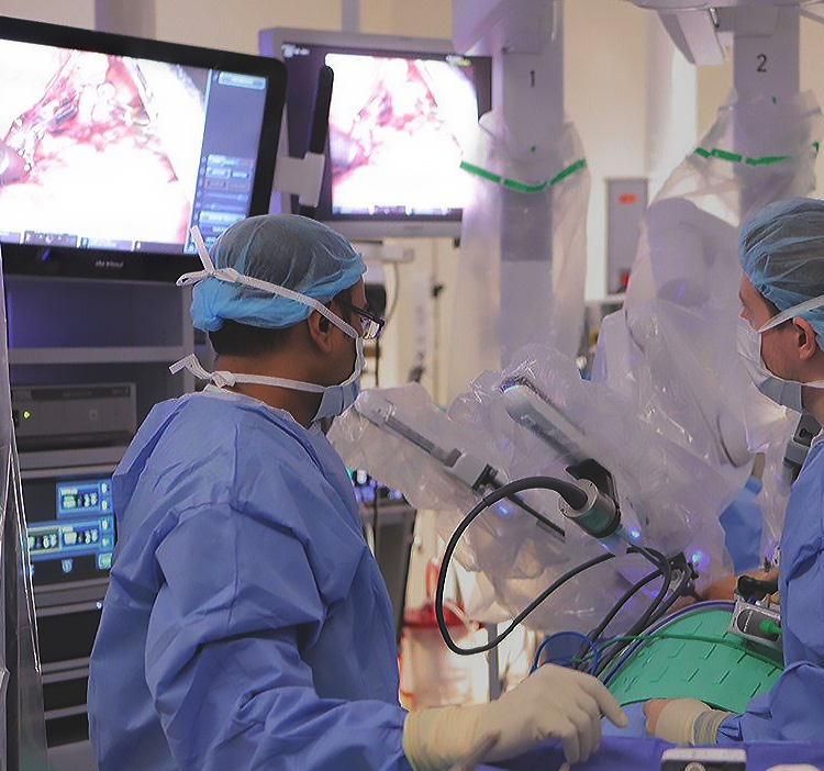
Some stomach cancer operations can be performed using laparoscopic or robot-assisted techniques through smaller incisions. These approaches may reduce certain postoperative burdens and facilitate recovery for appropriately selected patients. However, the choice between minimally invasive and open surgery depends on cancer extent, the complexity of reconstruction, patient factors, and the ability to safely achieve complete tumor removal and appropriate lymph node evaluation.
Chemotherapy and Systemic Treatment
Chemotherapy uses medications that circulate throughout the body to treat cancer cells. For many patients with localized but more advanced gastric adenocarcinoma, chemotherapy may be recommended before and after surgery, an approach called perioperative chemotherapy. Regimens such as FLOT may be considered for eligible patients. For unresectable or metastatic disease, chemotherapy may form the foundation of treatment and can be combined with other medications depending on tumor biology and the patient’s condition.
Radiation Therapy
Radiation therapy uses focused high-energy treatment to damage cancer cells. It may be recommended in selected circumstances, including certain postoperative situations or when treatment is needed to control bleeding, pain, or obstruction. Radiation may also be combined with chemotherapy for some patients. Its role is determined by the tumor’s location, prior treatment, and individual clinical circumstances rather than being routinely necessary for every stomach cancer diagnosis.
Targeted Therapy and Immunotherapy
Molecular and biomarker testing can identify specific characteristics of stomach cancer that may guide treatment, particularly when the disease is advanced or metastatic. Important biomarkers include HER2, PD-L1, mismatch repair deficiency or microsatellite instability (dMMR/MSI-H), and CLDN18.2. Depending on the findings, selected patients may benefit from HER2-directed therapies, immune checkpoint inhibitors, or other targeted treatments combined with chemotherapy. These medications are not appropriate for every tumor, making individualized molecular evaluation an important part of treatment planning.
Supportive Care and Clinical Trials
Nutrition and symptom management are essential components of stomach cancer care, particularly for patients experiencing weight loss, difficulty eating, anemia, or changes in digestion after treatment. Supportive care may involve nutrition specialists, pain management, psychosocial services, and procedures to address bleeding or obstruction. Clinical trials may also provide opportunities to consider investigational treatments when an appropriate study is available and eligibility requirements are met.
Learn More About Stomach Cancer Treatment
For additional information about surgical procedures, chemotherapy, radiation therapy, and other treatment options, visit our Stomach Cancer Treatment page.

At Saint John’s Cancer Institute, our multidisciplinary approach brings together specialists in surgical oncology, medical oncology, radiation oncology, gastroenterology, diagnostic imaging, and supportive care to evaluate complex stomach cancers and develop individualized treatment plans.
Through coordinated expertise, personalized treatment planning, and ongoing support, we help patients and families understand their diagnosis, explore available therapies, and make informed decisions about the next steps in their care.
If you have questions about gastric symptoms, stomach cancer, or treatment, please call today. Click here to request an appointment.