A high-risk breast lesion is an abnormal finding in breast tissue that is not necessarily cancer but may be associated with an increased risk of developing breast cancer or with a greater chance of finding a more significant abnormality nearby. These lesions are often discovered through breast imaging and biopsy, and the exact diagnosis helps your breast care team determine whether additional evaluation, surgical removal or ongoing monitoring is appropriate.
What Is a High-Risk Breast Lesion?
High-risk breast lesions are a group of tissue changes identified by a pathologist after a breast biopsy. The term does not mean that breast cancer is present or that cancer will inevitably develop. Instead, different high-risk lesions provide different information about the tissue that was sampled and a person’s future breast cancer risk.
Some lesions contain abnormal cells associated with an increased risk of developing breast cancer over time. Others are important because a needle biopsy samples only part of an area seen on imaging, and additional tissue may occasionally reveal a more significant abnormality. This is why the pathology result is considered together with the mammogram, ultrasound, MRI or other imaging that led to the biopsy.
Common High-Risk Breast Lesions
The significance of a high-risk lesion depends on the type of abnormality, whether atypical cells are present, how the lesion appeared on imaging and how well the biopsy findings explain the imaging abnormality. These factors help determine whether the area can be monitored or whether additional tissue should be examined.
Atypical Ductal Hyperplasia (ADH)
Atypical ductal hyperplasia is an abnormal growth of cells within the breast ducts. The cells have some features similar to those seen in ductal carcinoma in situ (DCIS), but ADH is not breast cancer. It is associated with an increased future risk of breast cancer and can sometimes be found near a more significant abnormality.
Because a needle biopsy examines only a portion of the area, surgical excision is commonly recommended after ADH is identified to determine whether nearby tissue contains DCIS or invasive cancer. In selected lower-risk situations, careful imaging surveillance may be considered after the imaging and pathology findings have been reviewed together.

Atypical Lobular Hyperplasia (ALH)
Atypical lobular hyperplasia involves abnormal cells within the lobules of the breast and is usually discovered incidentally during a biopsy performed for another imaging finding. ALH is not breast cancer, but it is considered a marker of increased future breast cancer risk.
When ALH is limited and the pathology findings adequately explain the imaging findings, ongoing surveillance may be appropriate. Additional tissue sampling or surgical excision may be recommended when the imaging and pathology do not correspond or when other concerning features are present.
Lobular Carcinoma In Situ (LCIS)
Despite the word “carcinoma” in its name, classic LCIS is generally considered a marker of increased breast cancer risk rather than an invasive breast cancer. Like ALH, it develops within the breast lobules and is often found incidentally during biopsy.
Management depends on the type of LCIS and the overall clinical findings. Classic LCIS may often be managed with surveillance when imaging and pathology are consistent, while non-classic forms such as pleomorphic or florid LCIS generally require additional evaluation and are more likely to be surgically removed.
Flat Epithelial Atypia (FEA)
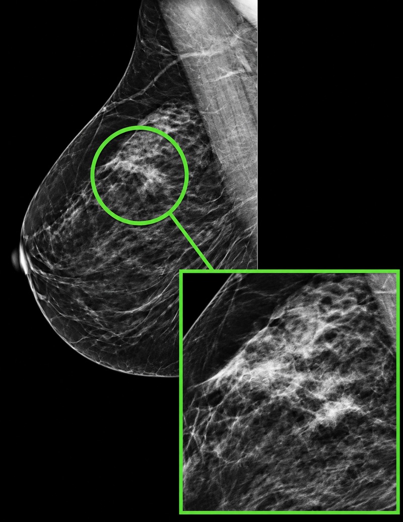
Flat epithelial atypia is a change involving cells lining the breast ducts and is usually discovered during a biopsy performed for an imaging abnormality such as calcifications. FEA itself is not breast cancer, but its significance depends on what else is present in the tissue and whether the biopsy adequately sampled the imaging finding.
Some patients with isolated FEA can be monitored rather than undergoing surgery. Additional sampling or surgical excision may be appropriate when other high-risk findings are present or when the pathology does not adequately explain the imaging abnormality.
Radial Scar and Complex Sclerosing Lesions
A radial scar is a benign breast lesion with a pattern of tissue that can sometimes resemble breast cancer on imaging. Despite its name, it is not a scar caused by an injury or previous surgery. Larger lesions with similar features may be called complex sclerosing lesions.
Management depends in part on whether atypical cells are present and whether the pathology findings are consistent with the imaging. Some lesions without atypia can be monitored, while surgical excision may be recommended when atypia or other concerning features are identified.
Intraductal Papilloma
An intraductal papilloma is a benign growth that develops within a milk duct and may sometimes cause clear or bloody nipple discharge. Papillomas may also be discovered through imaging or biopsy without causing symptoms.
The presence or absence of atypical cells is important when determining what happens next. A papilloma without atypia may sometimes be monitored when imaging and pathology are consistent, while papillomas containing atypia generally require additional evaluation and are more likely to be surgically removed.
What Happens After a High-Risk Breast Lesion Is Found?
A high-risk biopsy result is not interpreted in isolation. Your breast care team compares the pathology findings with the imaging abnormality that led to the biopsy. This process helps determine whether the tissue sampled provides a satisfactory explanation for what was seen on imaging.
Reviewing Imaging and Pathology Together
When the imaging and pathology findings are consistent, or concordant, the care team can determine whether surveillance or surgical removal is appropriate based on the specific lesion and your individual risk. When the findings do not correspond, additional sampling or surgical excision may be recommended because the original biopsy may not fully explain the abnormality seen on imaging.
When Is Surgical Excision Recommended?
Surgery is not automatically required for every high-risk breast lesion. The recommendation depends on the lesion type, whether atypia is present, the amount and quality of tissue obtained during biopsy, the imaging findings and the possibility that additional tissue could reveal DCIS or invasive breast cancer.
When excision is recommended, its immediate purpose is often to examine the area more completely and establish an accurate diagnosis. Your breast specialist can explain why surgery or surveillance is being considered in your particular situation.
Understanding Your Future Breast Cancer Risk
Managing the lesion that was found and understanding your future breast cancer risk are related, but they are not the same thing. Certain findings, particularly ADH, ALH and LCIS, can indicate an increased likelihood of developing breast cancer later in life. Surgically removing the area that was biopsied does not necessarily eliminate that underlying increase in risk.
Your care team may consider the biopsy finding together with your age, breast density, personal medical history, family history and other risk factors to develop an individualized plan. Depending on your overall risk, recommendations may include routine or enhanced breast imaging, clinical follow-up and discussion of medications that can reduce breast cancer risk for appropriate patients.
A high-risk breast lesion by itself does not necessarily mean that you have an inherited cancer predisposition. Genetic counseling or testing may be appropriate when your personal or family history suggests the possibility of an inherited cancer risk.
Learn more about Family History of Cancer and Genetics.
Moving Forward After a High-Risk Breast Biopsy
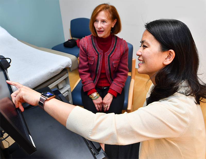
Learning that a breast biopsy contains a high-risk lesion can be confusing, particularly when the finding is described as abnormal but is not breast cancer. The most important next step is understanding exactly what was found, whether the pathology adequately explains the imaging abnormality and what that finding means for both the area that was biopsied and your future breast health.
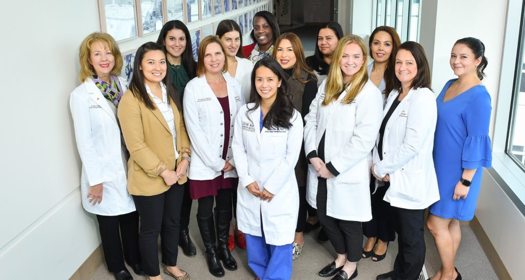
At the Margie Petersen Breast Center, breast imaging specialists, pathologists, surgeons and other members of the breast care team can help determine whether surveillance, additional biopsy, surgical excision or longer-term risk management is appropriate. Your plan should reflect the specific lesion and your individual circumstances rather than the label “high risk” alone.
Talk With Our Breast Clinic Team
The Breast Health Clinic can be reached at (310) 582-7209. If you have questions about a new breast symptom, need an evaluation or would like help determining your next step, call and a member of our team can assist you.