
Robotic Urologic Reconstruction
Urologic reconstruction restores or creates pathways for urine to move safely through the urinary system when normal drainage has been disrupted by obstruction, scar tissue, injury, previous surgery, radiation, cancer or other conditions. Depending on the problem, reconstructive surgery may repair a narrowed ureter, restore the connection between the kidney and ureter, or create a new way to store and pass urine after bladder removal.
Many reconstructive procedures can be performed using robotic-assisted surgery, which gives the surgeon magnified three-dimensional visualization and highly controlled instruments for working around the kidneys, ureters, bladder and surrounding structures. The robotic system is a surgical tool; the surgeon determines the type and extent of reconstruction based on the patient’s anatomy and the underlying condition.
Why Is Urinary Reconstruction Needed?

The urinary tract depends on open, functioning pathways to carry urine from the kidneys through the ureters to the bladder and out of the body. When one of these pathways becomes narrowed, blocked, damaged or must be surgically removed, urine may not drain normally. Depending on the location and severity of the problem, this can cause pain, infection, impaired kidney function or other complications.
Reconstructive surgery is designed to restore drainage while preserving healthy urinary tissue and kidney function whenever possible. When the bladder must be removed for cancer, reconstruction instead creates a new pathway for collecting and eliminating urine.
Reconstruction Is Individualized
There is no single reconstructive procedure for every urinary obstruction or injury. The appropriate approach depends on where the problem is located, its cause and extent, the condition of surrounding tissue, kidney and bladder function, previous surgery or radiation, and the patient’s overall health and treatment goals.
At Providence Saint John’s Health Center, reconstructive options may include repair of ureteral strictures, pyeloplasty for ureteropelvic junction (UPJ) obstruction and urinary reconstruction or diversion after bladder removal. The goal is to select the approach that restores urinary function while addressing the condition that made reconstruction necessary.
Robotic Repair of Ureteral Strictures
A ureteral stricture is a narrowing of one of the ureters, the tubes that carry urine from the kidneys to the bladder. Strictures can develop from scar tissue after previous surgery or injury, radiation therapy, inflammation and other conditions that affect the ureter. In some patients, nearby disease such as endometriosis or cancer can also narrow or obstruct the ureter.
When a stricture significantly interferes with urine flow, pressure can build above the narrowed area and affect the kidney. Depending on the severity and duration of the obstruction, patients may experience pain, recurrent urinary tract infections or progressive loss of kidney function. Some strictures cause few symptoms and are discovered during imaging or evaluation of kidney function.
How Are Ureteral Strictures Evaluated?
Before recommending reconstruction, the urologist determines where the stricture is located, how much of the ureter is affected and how well the kidney is functioning. Imaging and other diagnostic studies can help define the obstruction and determine whether urine is draining adequately from the kidney.
The cause of the stricture is also important. Previous abdominal or pelvic surgery, radiation, inflammation and other treatments can affect the condition and blood supply of the surrounding tissue and influence which reconstructive approach is most appropriate.
How Does Ureteral Reconstruction Work?
The goal of ureteral reconstruction is to bypass, remove or repair the narrowed portion of the ureter and restore a durable pathway for urine to drain from the kidney to the bladder. The specific technique depends on the location and length of the stricture and the amount of healthy ureter and surrounding tissue available for reconstruction.
For shorter strictures, the affected segment may be removed and healthy ends of the ureter reconnected. Strictures closer to the bladder may be treated by reconnecting the ureter to the bladder, while more complex or longer areas of narrowing may require additional reconstructive techniques to bridge or enlarge the affected segment.
Ureteroplasty and Complex Ureteral Reconstruction
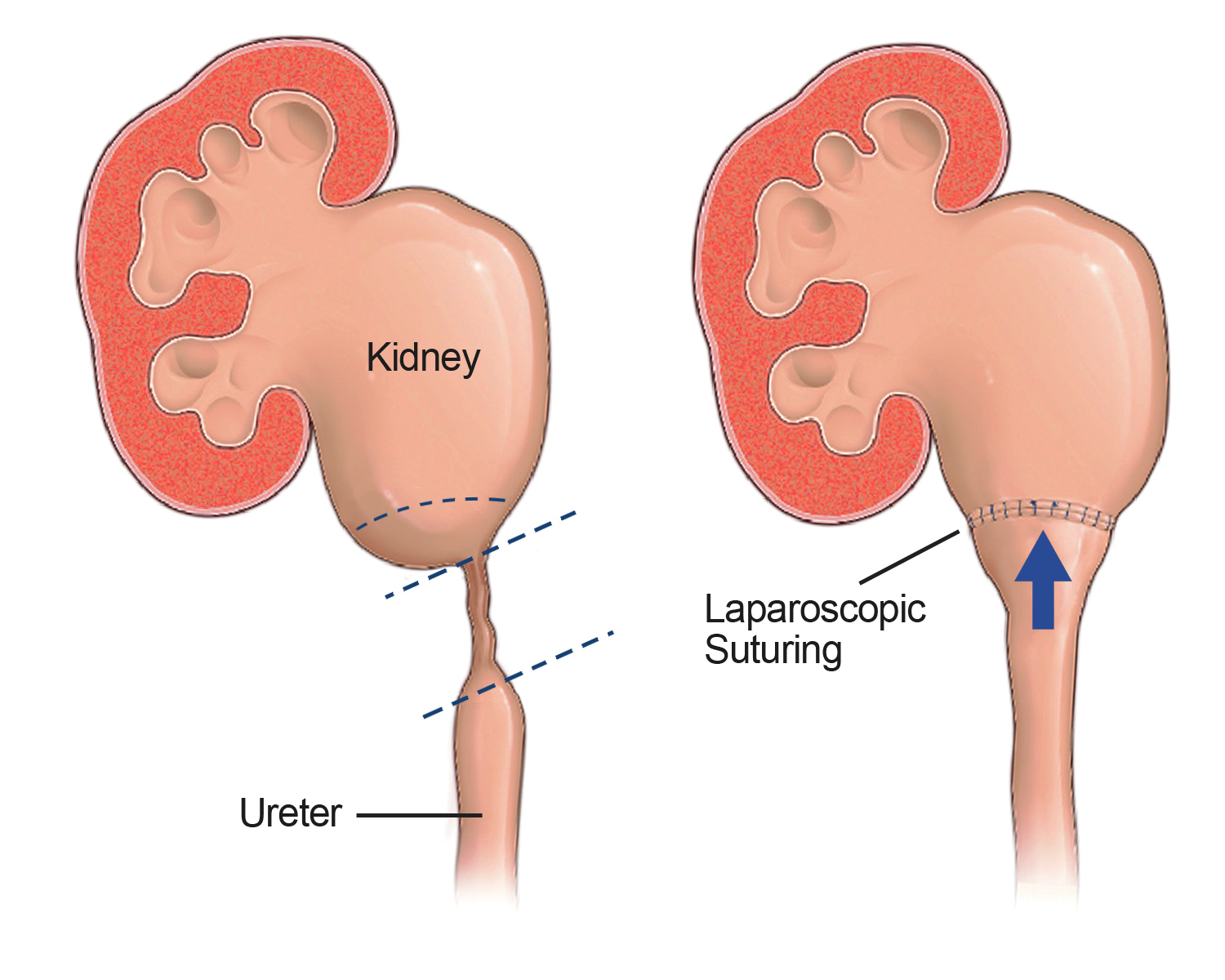
Ureteroplasty refers to reconstructive techniques used to repair a narrowed or damaged ureter while restoring urinary drainage. For selected longer or more complex strictures, tissue grafts may be used to widen or reconstruct the ureter when removing the entire narrowed segment and reconnecting the ends is not practical.
One contemporary approach uses a small graft of tissue from the inside of the cheek, called buccal mucosa, to reconstruct part of the ureter. This technique can provide another option for selected patients with complex strictures while allowing the surgeon to preserve healthy portions of the ureter.
Why Use Robotic-Assisted Surgery?
Robotic-assisted surgery allows the surgeon to perform many ureteral reconstructions through small incisions while viewing the operative area with magnified, three-dimensional visualization. Articulated instruments can help with the precise dissection and suturing required to reconstruct the ureter and surrounding urinary structures.
The robotic system does not determine which reconstruction should be performed. The surgeon selects and carries out the repair according to the patient’s anatomy, the characteristics of the stricture and the condition of the surrounding tissue.