Urinary obstruction can be the result of a variety of conditions. Our goal is to offer immediate and lasting relief, utilizing minimally invasive and best-practice techniques.
What is Urinary Obstruction?
Urinary obstruction, or urethral stricture, is the narrowing of the ureter tube. Urine can back up into the kidney and causes side and back pain, sometimes kidney infections, kidney stones or affect kidney function. Urologic reconstruction is the surgical repairing or recreation of a portion of urinary tract.
Causes of Ureteral stricture
- Any injury to the ureter during surgery can cause surgical scarring
- Can be caused by kidney stones, upper urinary tract inflammation, or a tumor. As scar tissue builds up in the ureter so does inflammation around the ureter
- Procedure to manage kidney stones
- Surgeries surrounding the ureters, such as gynecologic or vascular surgical procedures
- Radiation therapy for prostate cancer or other types of cancers
- Tumors
- Autoimmune conditions
- Congenital (present at birth)
Symptoms of Ureteral Stricture
- Side or back pain
- Feeling of fullness
- Blood in the urine
- Nausea
- Urinary tract infections
- Pain worse with increased fluids or alcohol
How do you test for Ureteral Stricture?
- Renal ultrasound
- CT scan
- MRI scan
- Renal Nuclear Medicine Scan
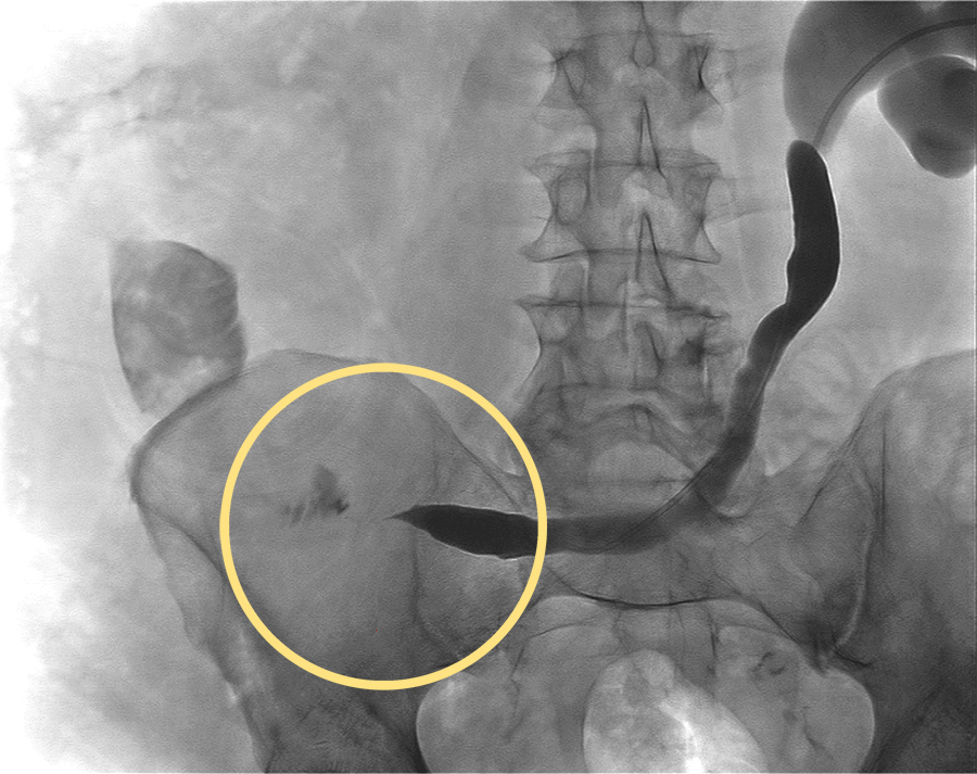
- Retrograde Ureteroscopy (Surgeon examine the ureter with a small camera to see why there is scarring)
Ureteropelvic Junction Obstruction (UPJ)
UPJ is a conditions where urine flow from the kidney into the ureter is blocked. The point of blockage is where the ureter (tube attached to kidney that drains urine to bladder) meets the collecting system or the portion of the kidney that collects the urine.
What Causes UPJ obstruction?
- Often a congenital condition which is present at birth
- Abnormal blood vessel over the ureter
- Scar tissue
- Infections
- Earlier treatments for a blockage
- Kidney stones
What are the signs and symptoms of UPJ obstruction?

The kidney can be damaged from the urine build up and this can cause:
- Abdominal mass
- Urinary tract infection
- Back or flank pain
- Bloody urine
- Kidney infection
- Vomiting
How is UPJ obstruction diagnosed?
- CT urogram (scan of both kidneys with contrast)
- Ultrasound
- Urine and blood test
- Nuclear scan of kidneys
- X-ray of kidneys, bladder, ureters
- Voiding cystourethrogram (X-ray of bladder while it is emptying)
Treatments for Urinary Obstruction

Robitic Surgery
When working near delicate and sensitive parts of the body, robotic surgery offers improved outcomes by enhancing the capabilities of the surgeon. The robot is controlled entirely by the urologist, replicating his or her delicate maneuvers precisely. Smaller incisions are needed with robotic surgery than traditional surgery, while the surgeon can also assess the surrounding areas and lymph nodes for signs of cancer spread (metastasis). Using a small camera, the surgeon views high resolution images on a video screen at the control console.
The da Vinci Surgical System translates the surgeon’s movements precisely using a smaller opening.
Benefits of robotic surgery include:
- Improved functional recovery
- Highly delicate surgical procedures and repairs can be made
- Reduced patient recovery time
- Shorter hospital stays
- Less post-surgical pain
After the cancerous area or organ has been removed, the robot is also used to repair areas of the body to restore function. They may include the use of tissue from the bladder, or bowel, to repair the small tubes that carry urine from the kidneys to the bladder. The robot is so precise that the surgeon can stitch very small tissues together in less space than by human hand. These maneuvers take place in such a small space that is helps reduce the patient’s recovery time. To help the ureter heal, a small flexible tube may be placed in the ureter to ensure it remains open while it is healing. The tube will remain in place 4 to 6 weeks. Your doctor will share additional information and care instructions before and after surgery, which will be essential to maximize your recovery.
Treatment of UPJ Obstruction: Robotic Pyleoplasty
Pyeloplasty has the best success for patients with UPJ obstruction. After laparoscopic pyeloplasty, there may be scarring in the abdomen area. Typically, the obstruction will not come back but that is possible.
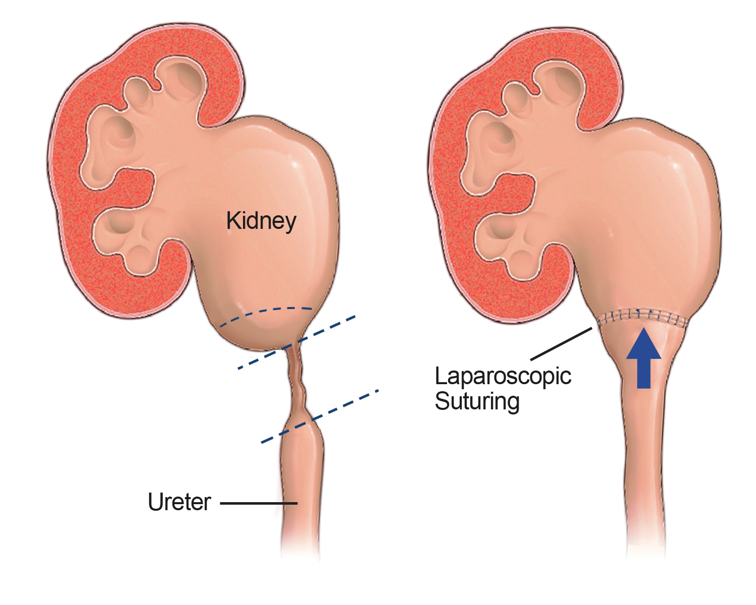
UPJ obstruction can be treated with surgery called a pyleoplasty. Sometimes this is treated very early in life to prevent future failure or injury of the kidney. When surgery is required, it is usually minimally invasive and referred to as pyeloplasty, which can takes a few hours. The portion of the ureter that is scarred or blocked by another vascular structure is typically removed or repaired. The normal ureter is then reattached to the renal pelvis of the kidney. A stent is left in the ureter until the repair is healed, which is usually 2 to 4 weeks.
What happens after pyeloplasty surgery?
- An overnight stay is expected for 1-2 days
- The ureter will still be swollen from the surgery, which may cause some discomfort
- A cystoscopy is performed 2 to 4 weeks later, which is the removal of the stent using a small flexible camera to remove the stent vial the bladder.
Risks for patients after surgery could be urinary tract infections or kidney stones.
Repair of Ureteral Strictures – Ureteroplasty
Ureteroplasty is a procedure and surgery that can fix narrowing of the ureters—tubes in the body that carry urine from the kidneys to the bladder. There are several reasons for ureteral stricture, including scar tissue that may have formed in a ureter. This can occur after trauma, operative injury to the ureter, or even have radiation treatment in that area of the body. Sometimes the ureter may be narrow from other medical issues such as endometriosis or cancer. When scarring occurs in the ureter, urine does not drain well. This is often painful and can lead to infection and long-term kidney damage.
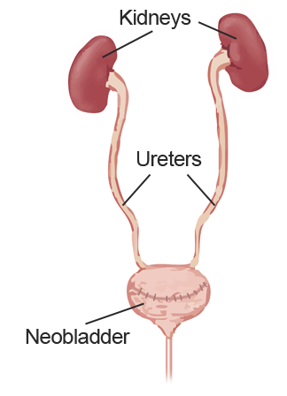
Reconstruction after Cystectomy
When bladder cancer forces the removal of the entire bladder, the surgeon creates another way for urine to be collected, stored and released from the body.
There are three ways this can be performed, while our physicians will help you decide which is best for you:
Artificial Bladder
A surgeon uses tissues from the intestines to create what is known as a neobladder. The neobladder is attached to the urethra and allows the patient to urinate normally. With this procedure, there is less blood loss and patients are able to return to normal physical activity much faster. Saint John’s surgeons are leaders in the field of this minimally invasive laparoscopic surgery and can ensure a highly successful outcome.
Urinary diversion with a bag outside the body
A surgeon uses intestinal tissue to create a path for urine to leave the body through an opening in the abdomen. This path is called a stoma, requiring a small opening in the abdomen and a bag that is positioned outside the body to collect urine.
Urinary diversion with a pouch inside the body
In this procedure, the surgeon uses intestinal tissue to create a reservoir called an Indiana Pouch and a path that leads out to the abdomen. The pouch collects urine inside the body. Several times a day, the patient uses a catheter (soft tube) to empty urine in the pouch. Providence Saint John’s specializes in this procedure.
Removal of the Bladder (After Cystectomy)
Often bladder cancer can require complete bladder removal, and for men, this usually means the prostate as well.
For women, this can involve removing the bladder and uterus, fallopian tubes, ovaries, and the top of the vaginal wall. When the bladder is removed, the surgeon must reconstruct the bladder so that the urine can pass from the kidney out of the body. There is no known artificial bladder available to patients so the surgeon must create one from intestine. Please See the Bladder cancer treatment page for more information.
If you have questions regarding urinary obstruction or treatments, please call today, or Click Here to make an appointment.